IMED3001 Study Guide - Midterm Guide: Phagocytosis, Alveolar Macrophage, Betamethasone

14 Dec 2020
School
Department
Course
Professor
Document Summary
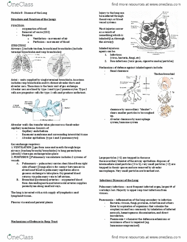
Refers to the cells across which gases must diffuse between the alveolar and capillary walls. The thickness of these layers varies from 0. 1 to 1. 5 m. Lining 95% of the surface of the alveoli. Edges of two alveolar cells overlap and are uniting by tight junctions preventing the leakage of blood/fluid from the alveolar septum to the alveolar space. Known as septal cells, lining only 5% of the alveolar surface. They are cuboidal cells that are interspersed among the type i pneumocytes. Surfactant reduces the surface tension in alveoli, preventing their collapse during expiration. They are progenitor cells for type i pneumocytes. Present within alveolar spaces (alveolar macrophage) and alveolar septum (septal macrophage). Slightly darker than type ii pneumocytes due to their content of dust and carbon. In alveolar space, they scavenging the alveolar surface to remove inhaled dust particles. In alveolar septum, they phagocytise rbcs that may enter the alveoli in heart failure.