BIO 263 Lecture 6: Anatomy Joints


JOINTS
Joints- two or more bones meet
- Direct contact, fiber tissue cartilage, fluid
- Immobile and slight mobile are more common on axial skeleton
- Classified on histological structure or range of motion
Synarthrosis- immovable
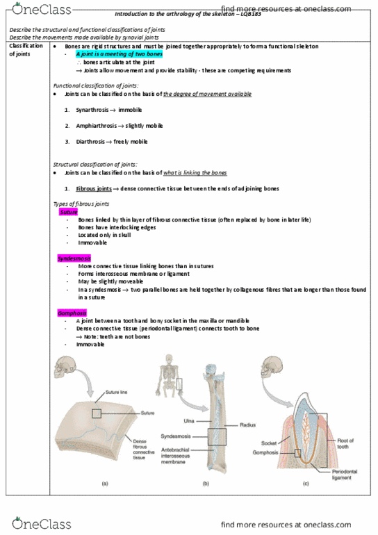
Fibrous:
1. Suture- fibrous joints of skull, edges are locked by fibrous connective tissue (sutural
ligament or membrane)
- Unossified remnants of embryonic mesenchymal membrane which bones developed
- Allow forces to be spread with minimal movement
2. Gomphosis- binds tooth to bony socket, periodontal ligament
Cartilaginous:
1. Synchondrosis- rigid, immovable
Bony Fusion
1. Synostosis- bones fuse from synchondrosis and boundary is gone
Amphiarthrosis-slight movement
Fibrous: connected by collagen fibers
1. Syndesmosis- bones are connected by a ligament (i.e. tibia and fibula)
Cartilaginous: connected by fibrous cartilage
1. Symphysis- separated by wedge/pad of fibrous cartilage (i.e. between pubic bones)
Diarthrosis- free movement
1. Synovial- ends of long bones
a. covered by articular cartilage, lack perichondrium/matrix has more fluid
b. Absorb shock and reduce friction
All Synovial have the same structure
A. Joint Capsule
B. Articular Cartilage
C. Joint Cavity with Synovial Fluid
D. Synovial membrane lining joint capsule
E. Accessory Structures
F. Sensory Nerves and blood vessels that supply interior/exterior of joint
Synovial Fluid Functions
1. Provide Lubrication: hyaluronan and lubricant with synovial fluid reduce friction
2. Nourish Chondrocytes: circulation is caused by joint movement, compression expansion
creates cycle for removal of waste and nourishment
3. Shock Absorber: cushion shock in joint that compress
Accessory Structures
find more resources at oneclass.com
find more resources at oneclass.com

1. Cartilage and Fat Pads
a. Meniscus: articular discs, cartilage that divide synovial cavity/flow of fluid/allow
shapes/restrictions
b. Fat Pads: around periphery, protect articular cartilage, fill spaces when moving
2. Ligaments
a. Accessory- support, strengthen, reinforce
b. Intrinsic- thickening of joint capsule
c. Extrinsic- separate from joint capsule
3. Tendons- pass across or around joint, limit range of motion
a. Sometimes a part of joint capsule for strength
4. Burase- small, fluid filled pockets in connective tissue
a. Filled with synovial fluid, lined by synovial membrane
b. Form where tendon and ligament rub against other tissues
c. Reduce friction, shock absorber
d. If developed in abdominal due to stress they are adventitious bursae
Strength vs Mobility
A joint cannot be highly mobile and very strong
Limiting Mobility and Reducing Injury:
1. Accessory ligaments and collagen fibers in joint capsule
2. Shapes of articulating surface prevent movement
3. Presence of other bones/bony processes/skeletal muscles/ fat pads
4. Tension in tendons
Angular Motions
1. Abduction- movement away from longitudinal axis (i.e. swing arms away from side),
appendicular
2. Adduction- bringing limbs back (i.e. unspreading toes), appendicular
3. Flexion- reduces angle, anterior posterior plane
4. Extension- increase angle between articulating elements, anterior posterior
a. Hyperextension: beyond normal limits
5. Circumduction- special type of angular motion, arms in a loop
Rotations
1. Left or Right Rotation- shaking head no
2. Internal Rotation- medial, towards body
3. External Rotation- lateral, away
4. Pronation- palm back
5. Supination- palm front
Special Movements
1. Eversion- turns foot sole out
2. Dorsiflexion- elevates toes when weight in heels
find more resources at oneclass.com
find more resources at oneclass.com
Document Summary
Classified on histological structure or range of motion. Immobile and slight mobile are more common on axial skeleton. Synarthrosis- immovable: suture- fibrous joints of skull, edges are locked by fibrous connective tissue (sutural ligament or membrane) Unossified remnants of embryonic mesenchymal membrane which bones developed. Allow forces to be spread with minimal movement. Gomphosis- binds tooth to bony socket, periodontal ligament. Bony fusion: synostosis- bones fuse from synchondrosis and boundary is gone. Amphiarthrosis-slight movement: syndesmosis- bones are connected by a ligament (i. e. tibia and fibula) Cartilaginous: connected by fibrous cartilage: symphysis- separated by wedge/pad of fibrous cartilage (i. e. between pubic bones, synovial- ends of long bones. Diarthrosis- free movement: covered by articular cartilage, lack perichondrium/matrix has more fluid, absorb shock and reduce friction. All synovial have the same structure: joint capsule, articular cartilage, joint cavity with synovial fluid, synovial membrane lining joint capsule, accessory structures, sensory nerves and blood vessels that supply interior/exterior of joint.