CAS BI 315 Lecture Notes - Lecture 69: Integrin, Vinblastine, Myosin Head


The Cytoskeleton (cont.)
- Specterin
- Different cells have similar linking proteins.
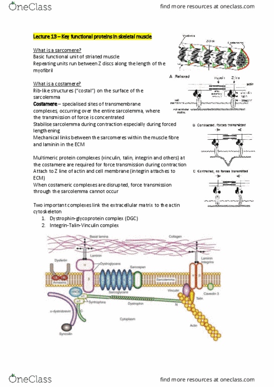
- Dystrophin (a calponin aka calcium binding protein) in muscle cells links actin
filaments to TM proteins in the plasma membrane, which link to the ECM
(Extracellular Matrix), help maintain cell stability during muscle contraction.
Dystrophin links actin to TM → TMs link to ECM → stable cell
- Muscular Dystrophy, is an X-linked inherited disease, results in
progressive degeneration of skeletal muscle.
- Dystrophin is absent (more severe) or abnormal (less severe) in patients
with Duchenne’s or Becker’s muscular dystrophy, respectively.
- Dystrophin doesn’t work and so it doesn’t connect the actin to the TMs.
- Actin, Myosin, and Cell Movement
- Muscle contraction: Actin filaments associate with myosin, a molecular motor, to
convert ATP to mechanical energy, generating force and movement.
- Striated muscles: Muscles composed of bundles of muscle fibers → Elongated
cells created by cell fusion during development (also why they’re multinucleated)
→ Long continuous stretches of cytoplasm. These cells are striated because of
the sarcomeres and distinct parts of the cell are held together by sarcomeres.
- Most of the cytoplasm is made up of myofibrils, which are composed of thick
filaments of myosin and thin filaments of actin. Together when they’re organized
into a contractile unit they’re called sarcomeres.
- Structure of a sarcomere
- Ends of each sarcomere defined by Z Disc
- Actin filaments are attached to Z disc by their barbed end.
- M line is in the middle of a sarcomere
- Myosin filaments attach to the M line
- Myosin and actin are overlapping but not connected.
- Muscle contraction
- The sliding of actin and
myosin filaments past
each other
- Z-discs get closer
- Where does the force
come from?
-Myosin II = motor protein
that drives filament sliding
→ has two key domains:
1) Globular head region
(GHR) 2) Coil of two
a-helical tails (aCoil)
- GHR binds actin and aCoil
associate in parallel to form staggered arrays in thick filament (interacts

with other myosin filaments to make one thick myosin filament) 100s -
1000s Myosins
- ATP-binding and hydrolysis drives myosin-mediated filament sliding
- Myosin associated with an actin molecule naturally → ATP pushes
away myosin head → ATP is hydrolyzed and myosin head binds
to new actin mol. and Pi is let go → Adp is let go and Myosin head
returns to original position and slides the filament with it.
- Hundreds of myosins contract a sarcomere
- How do muscle fibers regulate WHEN to contract?
- Presence of Ca+2 → Contraction | Absence of Ca+2 → NO contraction
- Nerve impulses increase Ca+2 100-fold
- Low Ca+2 → Tropomyosin-Troponin complex mask the myosin binding
site on actin.
- Troponin is a protein complex (made up of three parts we don’t
need to know) and they move tropomyosin based on the presence
or absence of Ca+2. (Tropomyosin blocks myosin)
- High Ca+2 → Ca+2 binds Troponin → shifts the Tropomyosin-Troponin
→ myosin binding site is unmasked
- The sarcoplasmic reticulum (ER of muscle cells) releases Ca+2
into the cytosol.
- Ca+2 will bind to the troponin complex, causing an allosteric
change that will move tropomyosin and expose myosin binding
sites.
- Cytokinesis: division of a cell following mitosis
- A contractile ring of actin and myosin II is assembled by membrane-bound
myosin just beneath the plasma membrane. Contraction of the ring pinches the
cell in two.
- How do vesicles and organelles move around the cell?
Unconventional myosins (don’t form filaments) such as myosin I and V that can
move along actin filaments take these vesicles and organelles around.
- Remember! Myosin HEAD will always interact with Actin
- Fig 13.32 is Myosin I and 13.33 is Myosin 5 (THE CUTE ONE)
Document Summary
Dystrophin (a calponin aka calcium binding protein) in muscle cells links actin filaments to tm proteins in the plasma membrane, which link to the ecm (extracellular matrix), help maintain cell stability during muscle contraction. Dystrophin links actin to tm tms link to ecm stable cell. Muscular dystrophy, is an x-linked inherited disease, results in progressive degeneration of skeletal muscle. Dystrophin is absent (more severe) or abnormal (less severe) in patients with duchenne"s or becker"s muscular dystrophy, respectively. Dystrophin doesn"t work and so it doesn"t connect the actin to the tms. Muscle contraction: actin filaments associate with myosin, a molecular motor, to convert atp to mechanical energy, generating force and movement. Striated muscles: muscles composed of bundles of muscle fibers elongated cells created by cell fusion during development (also why they"re multinucleated) These cells are striated because of the sarcomeres and distinct parts of the cell are held together by sarcomeres.