BIOL130 Lecture Notes - Lecture 14: Chemotaxis, Tx Network, Paravertebral Ganglia

28 Jan 2013
School
Department
Course
Professor
Document Summary
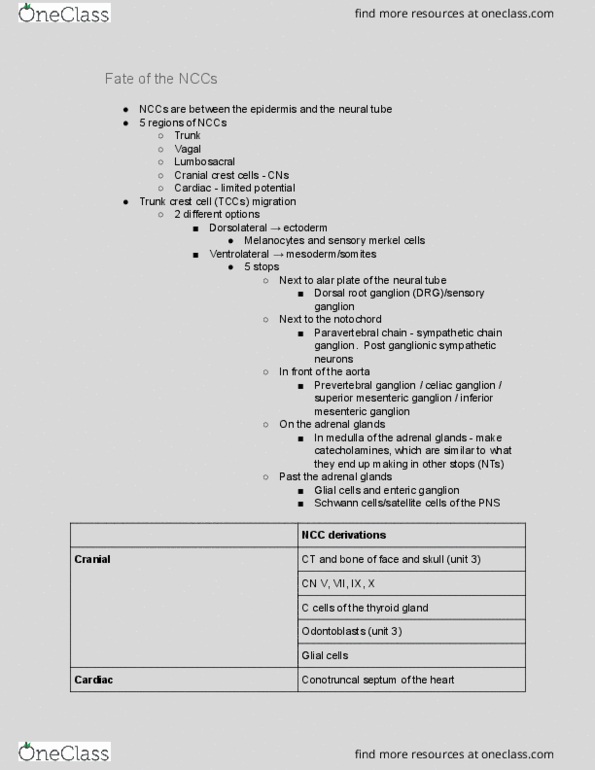
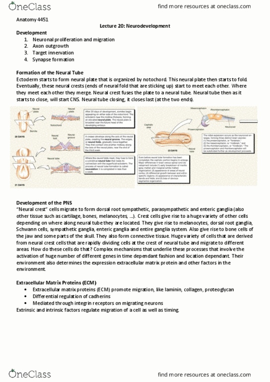
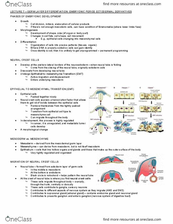
Neural crest cells: embryology and anatomy of nccs. Neural crest cells arise from dorsal ridge of neural fold and migrate to contribute to a wide variety of tissues. Major tissues formed by ncc include dorsal root ganglia, sympathetic chain ganglia, parasympathetic nervous system (e. g. enteric ganglia), melanocytes (see diagram p. 6) Combination of bmp (from neural tube) and wnt6 (ectoderm) signals cause. Ncc can migrate to anterior half of a somite, but not to posterior half due to repellent ecm; this gives rise to segmented pattern of the peripheral nervous system. Ncc cells migrate further to form drg and scg close to spinal cord (in contrast, parasympathetic components are close to the tissues they affect) Quail grafts in chicks can be used to track fate of nccs, since quail nuclei can be distinguished from chick nuclei. Nccs in trunk of body are a mixture of determined precursors and multipotent cells.