Health Sciences 4320A/B Lecture Notes - Lecture 7: Epithelial–Mesenchymal Transition, Neural Crest, Germ Layer

11 Feb 2020
School
Department
Course
Professor

LECTURE 7 – GERM LAYER DIFFERENTIATION: EMBRYONIC PERIOD ECTODERMAL DERIVATIVES
PHASES OF EMBRYONIC DEVELOPMENT
• Growth
Cell division, mitosis, elaboration of cellular products
If there’s not enough mesoderm cells, can have condition of Sirenomelia (where lower limbs fuse)
• Morphogenesis
Development of shape, size (of organ or body part)
Changes in cell fate, cell shape, cell movement
E.g., epithelial cells changing into mesenchymal cells
• Differentiation
Organization of cells into precise patterns (tissues, organs)
Where DNA is properly coded so cells can gain identity
Once identity is set, then it is unlikely to get unprogrammed – permanent programming
NEURAL CREST CELLS
• Develop at the postero-lateral borders of the neuroectoderm – when neural tube is folding
Come from the closing of the neural tube, originally ectoderm cells
• Dissociate from developing neural tube
• Undergo Epithelial-to-mesenchyme Transition (EMT)
Active migration and displacement
Enters underlying mesoderm
EPITHELIAL TO MESENCHYMAL TRANSITION (EMT)
• Epithelial cells
Packed together nicely
• Neural crest cells express a transcription factor that allows
them to get rid of bonds between the epithelial cells
Remove themselves from the tightly packed
arrangement
Transition from epithelial cell type to
mesenchyme cell
Can migrate throughout the body
• In development, this process is highly regulated
In cancer, it is unregulated, and metastatic tumor
cells develop
• A morphological change
MESODERM vs. MESENCHYME
• Mesoderm – derived from the mesodermal germ layer
• Mesenchyme – can derive from mesoderm, but is not itself mesoderm
• Epithelium – cells that line hollow organs and glands and those that make up the outer surface of the body
Very tightly regulated and organized
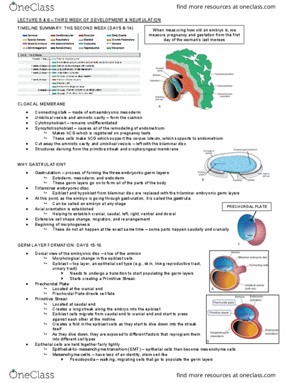
MIGRATION OF NEURAL CREST CELLS
• Neural tube – formed from ectoderm layer of germ cells
In the middle is mesoderm
At the bottom is endoderm
Black circle is notochord – helps pattern the neural tube
• At the crest of neural tube is where you find neural crest cells
These cells migrate throughout body – cranially,
through the trunk, ventrally or dorsally
These cells contribute to ganglia, sensory neurons
Contributes to different aspects of nervous system as they migrate (ANS and SNS)
Contributes to suprarenal gland (adrenal gland) – contains endocrine gland and neuronal gland
Contributes to preaortic ganglion and enteric ganglion (nervous system of digestive tract)

MIGRATION OF NEURAL CREST
• Cranial region – contributes to structures of the head and neck
• Trunk region – can either go dorsally or ventrally
1. Dorsal pathway through dermis – pigmented cells
(melanocytes)
2. Ventral pathway – through somites, helps populate
adrenal glands, sensory neurons, and glia
MIGRATION PATHWAYS OF CRANIAL NEURAL CREST CELLS
• Neural crest cells migrating cranially
They populate the pharyngeal arches
• Pharyngeal arches
Gill-like structures, odd looking
• Neural crest cells that migrate here populate the arches and contribute to the
cranial nerves V, VII, IX, X, and form the structures in the face and neck
Also contributes to the dentin in teeth
NEURAL CREST DERIVATIVES
• 4 Ganglia
Dorsal root ganglia of spinal nerves
Ganglia of V, VII, IV, X cranial nerves
Sympathetic and parasympathetic ganglia (autonomic nervous system)
Ganglion cell layer of the retina
• If neural crest cells take the dorsal route, they can become melanocytes
Pigmented cells
• Odontoblasts – produces the dentin of teeth
• Glia cells (Schwann cells) – creates myelin, which contributes to the neuron
Allows neuron to perform fast transmission of action potential
• Cranio-facial skeleton
• Adrenal Medulla
• Pia Mater and Arachnoid Mater (meninges) – tissue that lines the spinal cord
ECTODERMAL DERIVATIVES
• Anything that gives structures that maintain contact with the outside world – e.g., sensory structures, dermis
Central Nervous System
Peripheral Nervous System
Sensory epithelium (ears, nose, eyes)
Epidermis (hair, nails)
• Also derives:
Cutaneous glands – located in skin (e.g., sweat glands)
Mammary glands – used for nursing
Pituitary glands – hormones
Enamel of teeth
Lens of the eye
CLINICAL CORRELATES
Ectodermal Dysplasia
• So many different types of
ectoderm dysplasia’s
• Patchiness in hair, problems with teeth, very dry skin, tail growth
is uneven, some facial deformities
Document Summary
Lecture 7 germ layer differentiation: embryonic period ectodermal derivatives. Cell division, mitosis, elaboration of cellular products. If there"s not enough mesoderm cells, can have condition of sirenomelia (where lower limbs fuse: morphogenesis. Development of shape, size (of organ or body part) Changes in cell fate, cell shape, cell movement: differentiation. E. g. , epithelial cells changing into mesenchymal cells. Organization of cells into precise patterns (tissues, organs) Where dna is properly coded so cells can gain identity. Once identity is set, then it is unlikely to get unprogrammed permanent programming. Neural crest cells: develop at the postero-lateral borders of the neuroectoderm when neural tube is folding. Come from the closing of the neural tube, originally ectoderm cells: dissociate from developing neural tube, undergo epithelial-to-mesenchyme transition (emt) Epithelial to mesenchymal transition (emt: epithelial cells. Packed together nicely: neural crest cells express a transcription factor that allows them to get rid of bonds between the epithelial cells. Remove themselves from the tightly packed arrangement.