MEDRADSC 3C03 Lecture Notes - Lecture 19: Biliary Tract, Cholangiography, Endoscopic Retrograde Cholangiopancreatography

20 Aug 2020
School
Department
Course
Professor
Document Summary
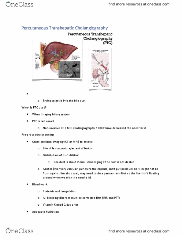
Looking at vasculature with use of contrast media. Needle inserted, in b/n the ribs, into the liver parenchyma into one of the vessels within the liver. When injecting contrast media into the biliary tract, we often see dilated vessels (bc of an obstruction to fluid movement) Sometimes it is hard stenting the area when we have dilated vessels. Dilation is normal, but could make a procedure more difficult to perform. Used to image the biliary system - considered a diagnostic procedure, just taking images. Invasive bc creating an artificial hole in the body. Non-invasive ct & mri cholangiography (still using cm to image flow through biliary system) have decreased the need for (diagnostics/exploratory): Ercp have decreased the need for ptc, now we are moving towards non-invasive procedures like ct/mri. Endoscope gains access into tree from esophagus, through sphincter of oddi in the duodenum.