PSYC 427 Lecture Notes - Lecture 15: Fastigial Nucleus, Dentate Nucleus, Pontine Nuclei

14 May 2018
School
Department
Course
Professor

PSYC 427 – LECTURE 15
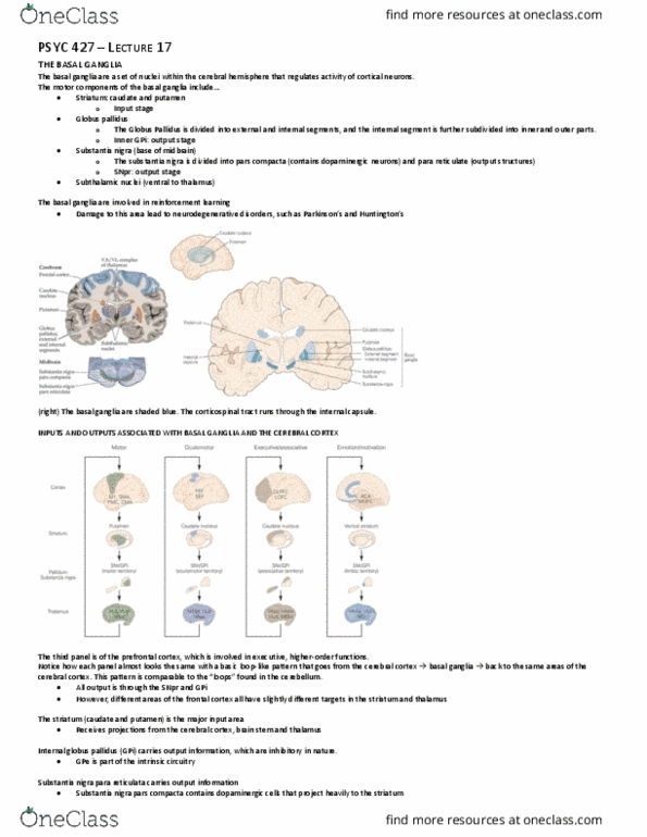
GROSS FEATURES OF CEREBELLUM
Dorsal view of the cerebellum
The cerebellum comprises an outer cover of gray matter (cerebellar cortex) and three pairs of deep nuclei (fastigial, anterior and posterior interpositus, and dentate)
• Dentate nucleus: most lateral
o Outputs to cerebral and prefrontal cortex
• Interpositus
o Outputs to cerebral cortex and the brainstem
• Fastigial nucleus: most interior
o Controls balance and eye movements
Two longitudinal furrows, that are most prominent ventrally, define an elevated midline ridge (vermis).
• The vermis runs the length of the cerebral cortex
Those with a cerebellar disorder have problems with movement coordination (ataxia).
• Difficulty learning motor skills
• Unable to adapt
The cerebellum has been around for a very long time- before the frontal cortices.
• Can be found in fish and birds
Outputs from the cerebellar cortex have targets in frontomotor areas, which in turn have outputs to areas of the cerebellum that originally sent out the signals
• These specific loops are also present in the prefrontal cortex, area 46 in particular.
Spinocerebellum: outputs and inputs relevant to the spinal cord
Nodulus and flocculus make up the base of the cerebellum, and are involved in vestibular function.
Superior cerebellar penduncle: carries efferent information from the deep nuclei to the thalamus
• Decussation (crossing) at the level of the inferior colliculus
• Purple: deep nuclei
• All afferent info entering the cerebral cortex goes out from the superior cerebellar penduncle
Middle cerebellar penduncle: carries pontine input from the cerebral cortex to the cerebellum
• Majority of afferent info goes through here (note that it is the biggest)
Inferior cerebellar penduncle: carries efferent info from the spinocerebellar tract (lower limb) and cuneocerebellar tract (upper limb) to ipsilateral cerebellum
• About half of the ascending signal that goes up from the cuneocerebellar cortex goes into the cerebellar cortex
There is crossing of information to create a consistent mapping between the left and right side of the brain.
• Info from the spinal cord remains on the ipsilateral side and crosses on its way to the cerebellar cortex
• Information from the periphery does’t ross
find more resources at oneclass.com
find more resources at oneclass.com

Frontal and parietal cortices are the main sources of info into the cerebellum.
• Note that the connections cross at midline
The pathway from frontal cortex to the inferior olive is important for adaptation
• Climbing fiber inputs have a powerful synaptic effect on purkinje neurons (main output neurons)
The Clarke nuclei is associated with the spinocerebellar pathway
Cotrol of upper li oveets are ediated iputs that…
1. Travel through the pontine nucleus
2. Cross at the midline
3. Enter the middle cerebellar penduncle
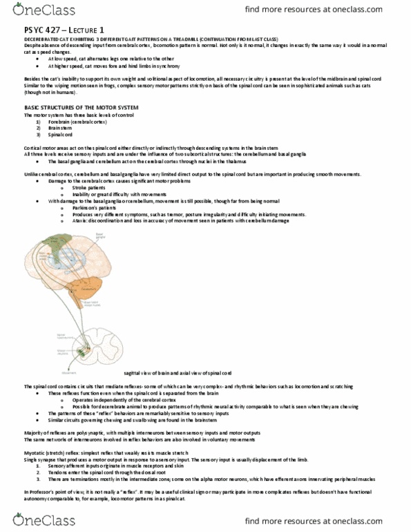
Projections from the cortex to the cerebellum
A retrograde tracer (WGA-HRP) was injected into pontine nuclei to identify cells in the parietal and frontal cortices that project to the pontine nucleus (origin).
• Dots: inputs to cerebellum (direct route)
Findings
1. Main inputs into the cerebellum come from the frontal cortex (areas 4 and 6)
2. There are also significant inputs from primary somatosensory cortex (areas 3, 1 and 2)
3. Note that there are almost no inputs from the auditory and visual cortices.
• Uneven sampling of cortical areas with direct outputs to the cerebellar cortex
find more resources at oneclass.com
find more resources at oneclass.com

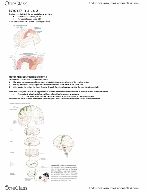
At the base of the cerebellum are output nuclei that project up to the cerebral cortex (purple)
• In a separate part of the cerebellum
1. Outputs go through the deep nucleus and superior penduncle and into the cortex via the ventrolateral thalamus
2. The superior colliculus, in turn, goes back into the inferior olive as a source of feedback for the cerebellar cortex
3. Outputs terminate on the contralateral thalamus and have targets in cerebral cortex
Outputs from the interpositus and dentate nuclei cross at midline
• Dentate above and interpositus below
Dum and Strick (2003) injected retrograde trans-neuronal transport herpes simplex virus (HSV1) into ereral orte to lael seod-order euros i detate
nucleus that project to cortex.
Post-injection survival time allowed retrograde transport of HSV1 from the injection site to first-order neurons in the thalamus, then retrograde transport to second-
order neurons in the dentate nucleus.
Finding: the dentate nucleus appears to be functionally divided into separate motor and non-motor domains
• The dentate nucleus is the main output source of input into the cerebral cortex
Additional survive time allowed for transport to third-order neurons in the putamen.
find more resources at oneclass.com
find more resources at oneclass.com
Document Summary
The cerebellum comprises an outer cover of gray matter (cerebellar cortex) and three pairs of deep nuclei (fastigial, anterior and posterior interpositus, and dentate) Two longitudinal furrows, that are most prominent ventrally, define an elevated midline ridge (vermis). The vermis runs the length of the cerebral cortex. Those with a cerebellar disorder have problems with movement coordination (ataxia). The cerebellum has been around for a very long time- before the frontal cortices. Outputs from the cerebellar cortex have targets in frontomotor areas, which in turn have outputs to areas of the cerebellum that originally sent out the signals. These specific loops are also present in the prefrontal cortex, area 46 in particular. Spinocerebellum: outputs and inputs relevant to the spinal cord. Nodulus and flocculus make up the base of the cerebellum, and are involved in vestibular function. Superior cerebellar penduncle: carries efferent information from the deep nuclei to the thalamus. Decussation (crossing) at the level of the inferior colliculus.