PSYC 427 Lecture Notes - Lecture 2: Ventral Posterolateral Nucleus, Cuneate Fasciculus, Alpha Motor Neuron

14 May 2018
School
Department
Course
Professor

PSYC 427 – LECTURE 2
The to suli that flak the postetral grus are the…
• Central sulcus: areas 4, 3a, 3b
• Postcentral sulcus: areas 2,5,7
In between the two sulci is area 1 on the gyrus itself
MOTOR AND SOMATOSENSORY CORTEX
DESCENDING LATERAL CORTICOSPINAL PATHWAY
1. The upper motor neurons of these tracts originate in the precentral gyrus of the cerebral cortex.
2. Fibers pass without synapsing all the way to their terminal destinations in the spinal cord
3. After leaving the cortex, the fibers descend through the internal capsule and into the pons then the medulla.
Most (about 75%) cross over to the opposite side, descend into the dorsolateral column to form the lateral corticospinal tract
• Terminates in lateral part of ventral horn, where the alpha motor neurons are
o The alpha motor neurons then send outputs to peripheral muscle, causing movement
The uncrossed fibers descend on the same (ipsilateral) side of the spinal cord to form the ventral corticospinal tract
find more resources at oneclass.com
find more resources at oneclass.com

ASCENDING DORSAL COLUMN-MEDIAL LEMNISCAL PATHWAY
1. Sensory inputs are sent to cell bodies of first-order neurons outside the spinal cord and in the dorsal root ganglion
2. Axons pass through the dorsal root
3. Aos for…
• Fasciculus gracilis: located medially; carries info from lower body
• Fasciculus cuneatus: located more laterally; carries info from upper body
4. The axons ascend ipsilaterally to the medulla
5. Cell bodies of second-order neurons form the gracile and cuneate nuclei
6. Axons cross over to form the medial lemniscus, ascending into the contralateral brainstem
** patha does’t ross oer util it is at the leel of the edulla **
7. Project into the ventral posterior lateral nucleus (VPL) of the thalamus
8. Third-order neurons send axons to the somatosensory cortex
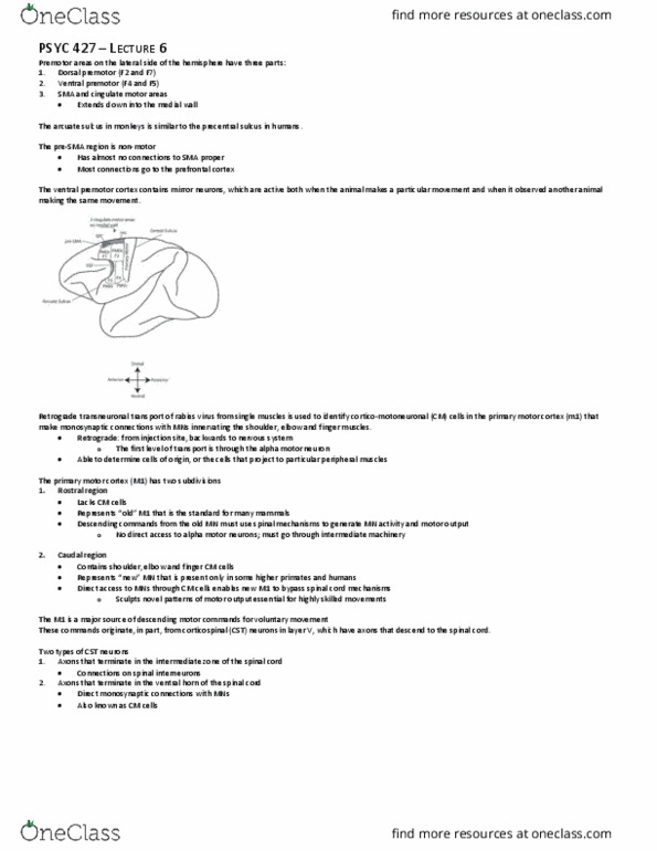
ROBOTIC COIL POSITIONING TMS EXPERIMENT
Optimal imaging system used to magnetically stimulate specific areas of the brain
• Enables recording of stimulations and corresponding movements
• Able to infer the pathway from the cortex to the periphery
The stimulating coil can be observed tracking the location of the brain in real time
• The coil is contralateral to the muscles that are being recorded using electrodes
• Advanced robotic technology allows adjustment of the coil to stimulate specific brain locations despite subtle movements made by the participant
• A stylus is run across landmarks on the brain to register the alignment
Professor’s understanding is that there are no uncrossed fibers that travel ipsilaterally all the way to the hand muscles
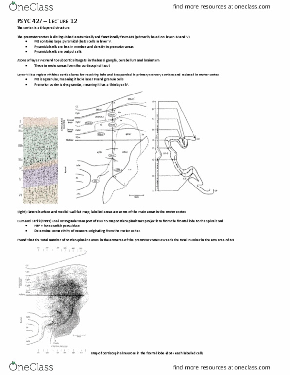
SEGMENTS OF THE SPINAL CORD
Below is a cross-sectional image of the spinal cord when injected with a tracer to the left primary motor and somatosensory cortices
• Anterograde tracer propagates from the injection site along the level of the axon
• Termination sites at the level of spinal cord are pictured as dots
o Right: contralateral
o Left: ipsilateral
Segments of the spinal cord from top (rostral) to bottom (caudal): low cervical, thoracic, lumbar and sacral
• The highest up is the low cervical area around the neck
• Two enlargements: cervical and lumbar
o Many fibers terminate here since there are projections to the arms and lower limbs
find more resources at oneclass.com
find more resources at oneclass.com
Document Summary
The t(cid:449)o sul(cid:272)i that fla(cid:374)k the post(cid:272)e(cid:374)tral g(cid:455)rus are the . In between the two sulci is area 1 on the gyrus itself. Most (about 75%) cross over to the opposite side, descend into the dorsolateral column to form the lateral corticospinal tract. The upper motor neurons of these tracts originate in the precentral gyrus of the cerebral cortex. Fibers pass without synapsing all the way to their terminal destinations in the spinal cord. After leaving the cortex, the fibers descend through the internal capsule and into the pons then the medulla. Terminates in lateral part of ventral horn, where the alpha motor neurons are. The alpha motor neurons then send outputs to peripheral muscle, causing movement. The uncrossed fibers descend on the same (ipsilateral) side of the spinal cord to form the ventral corticospinal tract. Sensory inputs are sent to cell bodies of first-order neurons outside the spinal cord and in the dorsal root ganglion.