LQB183 Lecture Notes - Lecture 4: Exocrine Gland, Abdomen, Ampulla

9 Aug 2018
School
Department
Course
Professor
Document Summary
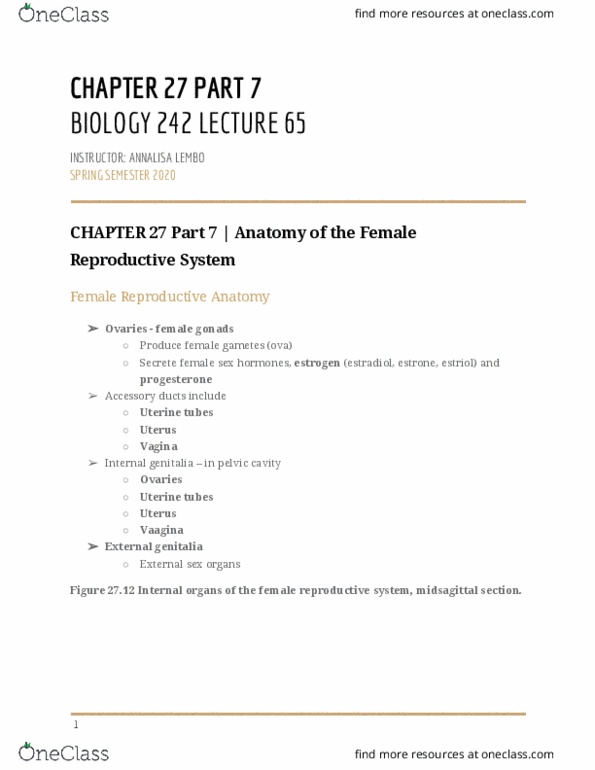
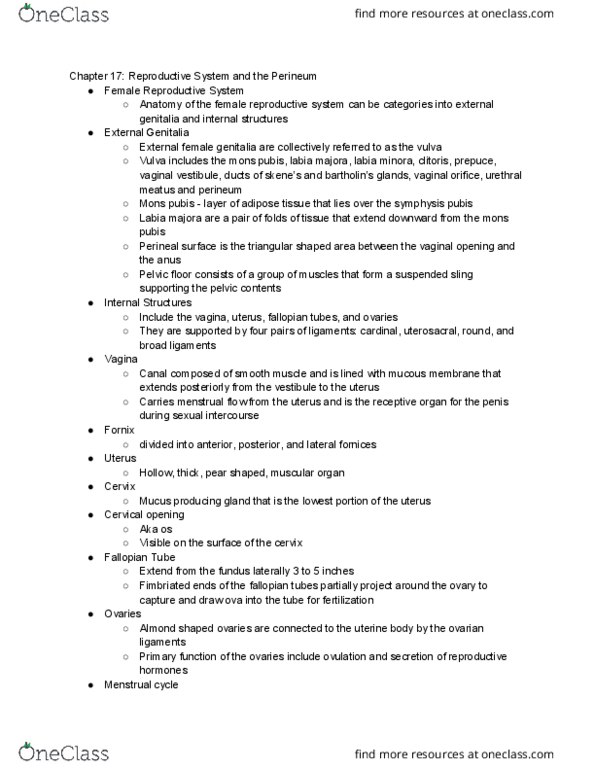
Describe and identify the macroscopic anatomy of the external and internal genitalia of the female reproductive system. The female reproductive system consists of the internal genitalia including the paired ovaries, paired uterine tubes, a single uterus, a single vagina and the vulva/external genitalia. Function of the female reproductive system: reception of spermatozoa. Provision of a suitable environment for the fertilisation of ova by spermatozoa and for the development of the embryo and foetus. Located in pelvic cavity and held in place by various ligaments: external genitalia (vulva) Females have two uterine (fallopian) tubes extending laterally from the uterus. Provide a route for sperm to reach an ovum. Transport oocyte (fertilised ova) from ovaries to uterus. Infundibulum funnel-shaped portion of tubes, close to ovary but open to pelvic cavity: ends have finger-like projections called fimbriae. Ampulla widest, longest portion of uterine tube. Isthmus short, narrow portion of uterine tube. Fertilisation normally occurs in the fallopian tubes (uterine tubes)