ANAT20006 Lecture Notes - Lecture 22: Thoracic Wall, Vertebra, Central Tendon Of Diaphragm

23 Jul 2018
School
Department
Course
Professor
Document Summary
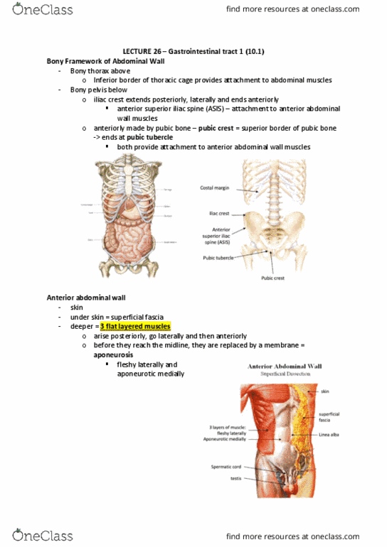
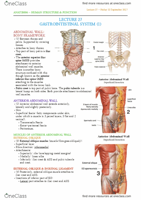
11th and 12th ri(cid:271) ha(cid:448)e free a(cid:374)terior e(cid:374)d. do(cid:374)"t atta(cid:272)h to ster(cid:374)u(cid:373) (cid:1012) (cid:1013) (cid:1005)(cid:1004) do(cid:374)"t atta(cid:272)h dire(cid:272)tly. Typical structure changes for top and bottom ribs. Costal groove is a pathway for neurovascular bundles. Between head of rib, inferior facet of vertebra above, superior facet of vertebra below and. Between transverse process and tubercle part of rib (smooth part) Superior bordered by sternum, 1st rib, t1, manubrim and costal cartilages. Covered by suprapleural membrane and partially closes it laterally. Inferior border by costal margin, t12, 12th rib and closed by diaphragm. Circumferential- muscle arises peripherally from inferior border and attaches to central tendon (cid:449)ithout (cid:271)o(cid:374)es. Aorta (cid:271)ehi(cid:374)d so that it is(cid:374)"t (cid:272)ha(cid:374)ged (cid:271)y (cid:272)o(cid:374)tra(cid:272)tio(cid:374)s. Phrenic= diaphragm (motor nerve muscles, nerves, arteries, veins. When contracts lifts ribs below up and out. When contract pulls rib in and increases pressure in thoracic cavity. Inner- not for respiration, run in diff directions.