ANAT20006 Lecture Notes - Lecture 27: Anterior Inferior Iliac Spine, Anterior Superior Iliac Spine, Abdominal External Oblique Muscle

12 Jun 2018
School
Department
Course
Professor

LECTURE 27
GASTROINTESTINAL SYSTEM (1)
ABDOMINAL WALL:
BONY FRAMEWORK
•(1) Between thorax and
pelvis. Supported by covering
tissues.
•Attaches to bony thorax.
•Top part of bony pelvis is iliac
crest.
•The anterior superior iliac
spine (ASIS) provides
attachment to anterior
abdominal wall muscles.
There is another bony
structure confused with this
though known as the anterior
inferior iliac spine (AIIS)
attaching to the muscles
associated with the lower limb.
•Pubic crest is top part of pubic bone. The pubic tubercle is a
lateral bump on both sides. Both provide attachment to abdominal
wall muscles.
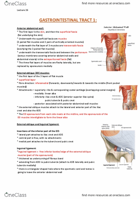
ANTERIOR ABDOMINAL WALL
•(2) Anterior abdominal wall extends anteriorly,
laterally and slightly posteriorly.
•Skin
•Superficial fascia: fatty components under skin,
under which is muscle in 3 paired layers, 3 flat and 2
vertical).
•Transversalis Fascia
•Extra-peritoneal fascia
•Peritoneum.
MUSCLES OF ANTERIOR ABDOMINAL WALL
EXTERNAL OBLIQUE
•(3) External oblique muscles (muscle fibre goes obliquely):
•Superficial layer
•Fibre direction: inferomedial
•Attachments:
•Superiorly: ribs (overlapping costal margins)
•Medially: linear alba
•Inferiorly: iliac crest & ASIS and pubic tubercle
and crest.
EXTERNAL OBLIQUE & INGUINAL LIGAMENT
•(4) Posteriorly, external oblique muscle attaches to
iliac crest and ASIS.
•Insertions of inferior part of EO:
•Lateral part attaches to iliac crest and ASIS
Lecture 27 - Friday 22 September 2017
ANAT20006 - HUMAN STRUCTURE & FUNCTION

•Central part is free with no attachments
•Medial part attaches to public tubercle and pubic crest.
•Inguinal ligament: named as it is in inguinal area
•Free inferior border of EO
•Thickened as an under-curving fibrous band curving
inwards
•Extending from ASIS to pubic tubercle
MUSCLES OF ANTERIOR ABDOMINAL WALL
INTERNAL OBLIQUE
•(5) Arise posteriorly.
•Internal oblique (IO) muscles:
•Intermediate layer
•Fibre direction: Superomedial
•Lowermost fibres: directly arise from lateral half of inguinal
ligament.
•Arise from lateral part (about 2/3) inguinal ligament
•Arch up and then downwards and then insert into pubic
crest via a conjoint tendon.
•Replaced by abdominal muscle aponeuroses fusion forming
linea alba, a line running down the midline from xiphoid
process to the pubic symphysis. Name = ‘white line’ which
it is, due to collagen. Separates left and right rectus
abdominus muscles (slide 8 details). Divides a 6 pack in
half.
•Superiorly attach to costal margin.
TRANSVERSUS ABDOMINUS
•(6) Fibre direction goes horizontally.
•Transverse Abdominus (TA) muscle:
•Innermost layer
•Fibre direction: transverse
•Lowermost fibres:
•Arise from lateral part (1/3) inguinal ligament
•Arch up and then downwards and then insert
into pubic crest via a single conjoint tendon.
•Conjoint tendon: When 2 structures insert into a bony
compartment via 1 tendon.
Lecture 27 - Friday 22 September 2017
ANAT20006 - HUMAN STRUCTURE & FUNCTION
Document Summary
Supported by covering tissues: attaches to bony thorax, top part of bony pelvis is iliac crest, the anterior superior iliac spine (asis) provides attachment to anterior abdominal wall muscles. There is another bony structure confused with this though known as the anterior inferior iliac spine (aiis) attaching to the muscles associated with the lower limb: pubic crest is top part of pubic bone. The pubic tubercle is a lateral bump on both sides. Anterior abdominal wall: (2) anterior abdominal wall extends anteriorly, laterally and slightly posteriorly, skin, superficial fascia: fatty components under skin, under which is muscle in 3 paired layers, 3 flat and 2 vertical), transversalis fascia, extra-peritoneal fascia, peritoneum. External oblique: (3) external oblique muscles (muscle fibre goes obliquely), superficial layer, fibre direction: inferomedial, attachments, superiorly: ribs (overlapping costal margins, medially: linear alba, inferiorly: iliac crest & asis and pubic tubercle and crest.