ANAT20006 Lecture Notes - Lecture 24: Pulmonary Pleurae, Thoracic Cavity, Rib Cage

12 Jun 2018
School
Department
Course
Professor

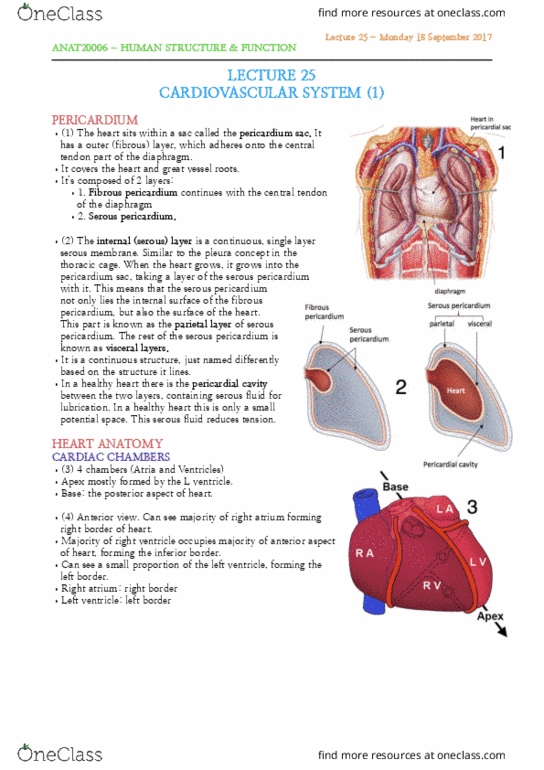
LECTURE 24
LOWER RESPIRATORY TRACT
THORACIC CAVITY
•(1) The thoracic cavity is divided into 3
major compartments; the left and right
pulmonary cavities and the mediastinum.
•The left and right lungs live in the left and
right pulmonary cavities, respectively.
•Anatomically, the lungs are separated
from each other.
•The mediastinum is basically everything
that’s not the lungs. It separates the lungs.
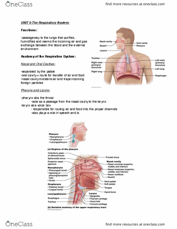
PLEURA
•(2) Lung surface covered by a continuous serous
membrane called pleura. The pleura covers the
surface of the lungs but once it gets to the lung
root, it reflects away and lines the mediastinum.
Thus it lines the wall of the thoracic cage.
•(3) Each lung is like a tree growing from the
mediastinum. This means, anatomically, they still
have to connect to the mediastinum in the middle,
by the lung root.
•The pleura is divided into 2 parts: visceral (lining
lungs) and parietal pleura (lining wall of cavity).
There is a pleural cavity (potential space) between
the two layers of pleural membrane containing a
few mls of serous pleural fluid, allowing friction free lung movement.
Lecture 24 - Friday 15 September 2017
ANAT20006 - HUMAN STRUCTURE & FUNCTION

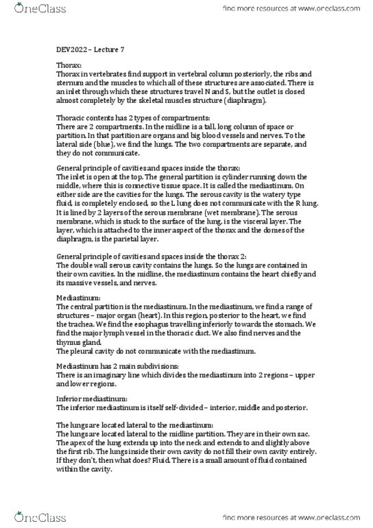
PARIETAL PLEURA
•(4) Divided into 4 parts.
•Cervical pleura (lining the cervical
extension of the pleural cavity)
•Costal pleura (related to the ribs and
intercostal space)
•Mediastinal pleura (covering the
mediastinum)
•Diaphragmatic pleura (covering the
diaphragm)
•(5) If we want to section a lung away from
the mediastinum we have to section a lung
root.
PULMONARY LIGAMENT
•(6) Shiny surface is visceral layer of pleura.
•The pleural sleeve surrounding the lung roots is very loose. Therefore
it hangs down to form a double folded structure inferior to the lung
root. Doesn’t attach to any bony structure or muscle. This is the
pulmonary ligament. It represents continuity between the parietal and
visceral pleurae.
PLEURAL NERVE INNERVATION
•(7) Visceral and parietal layer receive distinct nerve and vascular
supply.
•Lining tissues share the same nerve supply as the structure they
cover:
•Visceral pleurae receive autonomic nerve innervation
•Parietal pleurae receive somatic nerve innervation
Lecture 24 - Friday 15 September 2017
ANAT20006 - HUMAN STRUCTURE & FUNCTION
Document Summary
Pleura: (2) lung surface covered by a continuous serous membrane called pleura. The pleura covers the surface of the lungs but once it gets to the lung root, it reflects away and lines the mediastinum. Thus it lines the wall of the thoracic cage: (3) each lung is like a tree growing from the mediastinum. This means, anatomically, they still have to connect to the mediastinum in the middle, by the lung root: the pleura is divided into 2 parts: visceral (lining lungs) and parietal pleura (lining wall of cavity). There is a pleural cavity (potential space) between the two layers of pleural membrane containing a few mls of serous pleural fluid, allowing friction free lung movement. Pulmonary ligament: (6) shiny surface is visceral layer of pleura, the pleural sleeve surrounding the lung roots is very loose. Therefore it hangs down to form a double folded structure inferior to the lung root. Doesn"t attach to any bony structure or muscle.