ANAT20006 Lecture Notes - Lecture 25: Serous Membrane, Pericardium, Rib Cage

12 Jun 2018
School
Department
Course
Professor

LECTURE 25
CARDIOVASCULAR SYSTEM (1)
PERICARDIUM
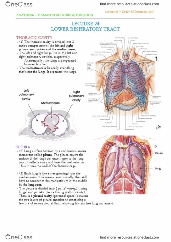
•(1) The heart sits within a sac called the pericardium sac. It
has a outer (fibrous) layer, which adheres onto the central
tendon part of the diaphragm.
•It covers the heart and great vessel roots.
•It’s composed of 2 layers:
•1. Fibrous pericardium continues with the central tendon
of the diaphragm
•2. Serous pericardium.
•(2) The internal (serous) layer is a continuous, single layer
serous membrane. Similar to the pleura concept in the
thoracic cage. When the heart grows, it grows into the
pericardium sac, taking a layer of the serous pericardium
with it. This means that the serous pericardium
not only lies the internal surface of the fibrous
pericardium, but also the surface of the heart.
This part is known as the parietal layer of serous
pericardium. The rest of the serous pericardium is
known as visceral layers.
•It is a continuous structure, just named differently
based on the structure it lines.
•In a healthy heart there is the pericardial cavity
between the two layers, containing serous fluid for
lubrication. In a healthy heart this is only a small
potential space. This serous fluid reduces tension.
HEART ANATOMY
CARDIAC CHAMBERS
•(3) 4 chambers (Atria and Ventricles)
•Apex mostly formed by the L ventricle.
•Base: the posterior aspect of heart.
•(4) Anterior view. Can see majority of right atrium forming
right border of heart.
•Majority of right ventricle occupies majority of anterior aspect
of heart, forming the inferior border.
•Can see a small proportion of the left ventricle, forming the
left border.
•Right atrium: right border
•Left ventricle: left border
Lecture 25 - Monday 18 September 2017
ANAT20006 - HUMAN STRUCTURE & FUNCTION

SURFACE ANATOMY OF THE
HEART
ANTERIOR VIEW
•(5) Can see right atrium appendage
and left atrium appendage at the front
of the heart.
•Sulcus (groove):
•Atrioventricular (coronary) sulcus
between right atrium and right
ventricle, appearing at the front.
So it is known as anterior
atrioventricular sulcus.
•Between the right and left
ventricles there is an
interventricular sulcus. Also
anterior because at front of heart.
•Atrium appendage (auricle)
POSTERIOR VIEW
•(6) Can see majority of left atrium at
back of heart. Can see a small proportion
of right atrium forming the right border.
•So majority of right chambers are at the
front but can still be seen at the back.
•The atrioventricular groove becomes
posterior once it runs to the back of the
heart.
RIGHT ATRIUM
•(7) Thin walled. Not pumps. Receiving
chambers. Majority of internal surface is
smooth, except for anterior wall which is
covered by musculi pectinati.
•Everything below the diaphragm drained
by the inferior vena cavae, Everything
above the diaphragm apart from heart
drained by superior vena cavae.
•Near the valves of the IVC is the
coronary sinus, which drains the heart to
the right atrium.
•In the posterior wall of the right atrium
there is a thumb print looking thing
called the fossa ovalis. In a developing
Lecture 25 - Monday 18 September 2017
ANAT20006 - HUMAN STRUCTURE & FUNCTION
Document Summary
Pericardium: (1) the heart sits within a sac called the pericardium sac. It has a outer (fibrous) layer, which adheres onto the central tendon part of the diaphragm: it covers the heart and great vessel roots, it"s composed of 2 layers, 1. Fibrous pericardium continues with the central tendon of the diaphragm: 2. Serous pericardium: (2) the internal (serous) layer is a continuous, single layer serous membrane. Similar to the pleura concept in the thoracic cage. When the heart grows, it grows into the pericardium sac, taking a layer of the serous pericardium with it. This means that the serous pericardium not only lies the internal surface of the fibrous pericardium, but also the surface of the heart. This part is known as the parietal layer of serous pericardium. In a healthy heart this is only a small potential space.