ANAT20006 Lecture Notes - Lecture 4: Yolk Sac, Gastrointestinal Tract, Allantois

12 Jun 2018
School
Department
Course
Professor

LECTURE 4
EMBRYOLOGY (3)
LECTURE 3:
•
ENDODERM
•(3) Important for inducing formation of mesodermal organs (Eg. heart, blood vessels)
•Forms lining of digestive tract
•Digestive tract: whole length of the body
•Buds off liver, gall bladder, pancreas, lungs
•Cranial part (anterior of lungs): pharynx
•Epithelial pockets of pharynx: thyroid, thymus, and parathyroid
BODY FOLDING
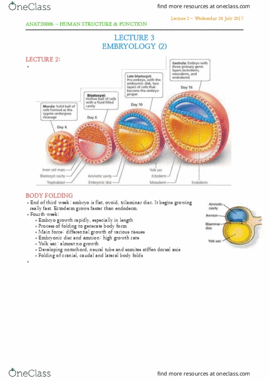
•(4) End of third week: embryo is flat, ovoid, trilaminar disc
•Fourth week:
•Embryo growth rapidly, especially in length
•Process of folding to generate body form
•Main force: differential growth of various tissues
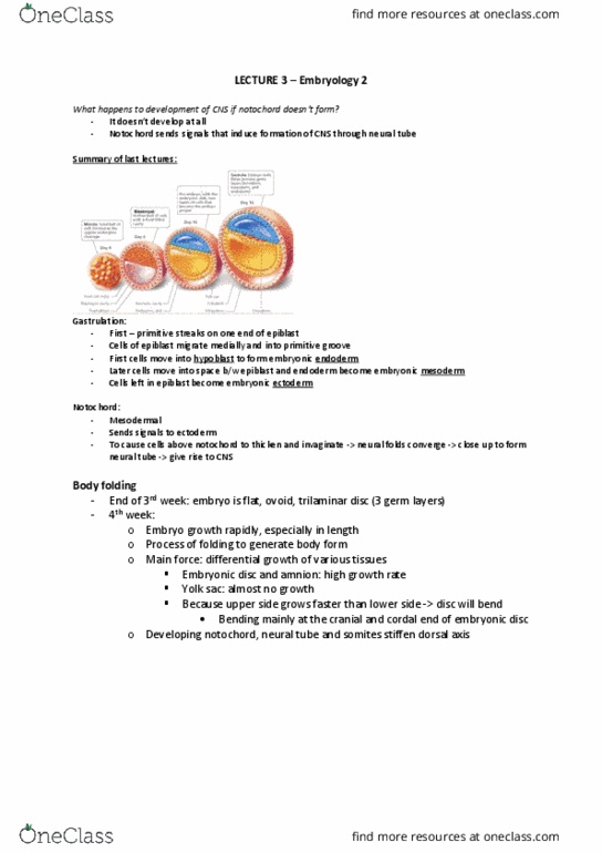
•Embryonic disc and amnion: high growth rate
•Yolk sac: almost no growth
•Folding of cranial, caudal and lateral body folds
•Body folding is folding of the embryo due to faster growth in one area than another.
•(5) connection to the yolk sac narrows more and more until it is only the stalk:
Lecture 4 - Monday 31 July 2017
ANAT20006 - HUMAN STRUCTURE & FUNCTION

•Transverse section: in most places there
is little bits that fuse together on the
sides.
•(6) animation 1: yolk sac gets smaller.
•(6) animation 2) allantois is endodermal.
As it gets smaller we can distinguish
different areas of the gut.
•(7) transverse section. Via body folding
we can see the sides come together to
fuse and we end up
with a gut tube lined
on the inside by
endoderm and on the
outside by mesoderm.
•Lateral folding of
the embryo
completes the gut
tube
•Mesodermal layer
of the gut tube
from splanchnic
mesoderm
•Somatic
mesoderm lines
body cavity.
SEPTUM
TRANSVERSUM
•(8) Mesodermal origin.
•When the head folds in, some of the mesoderm
gets pulled into the embryo and we can see the
heart forming. It really cuts the coelom size in
half.
•Separating coelom into thoracic and abdominal cavities
•Develops into part of diaphragm and ventral mesentery
of stomach and duodenum
ALLANTOIS
•(9) allantois: important for gas and nutrition exchange
and excretion within the embryo.
GUT DEVELOPMENT
•(10) Development: can distinguish 3 parts,
which are defined by their blood supply.
•Primitive gut develops at beginning of forth week
•Closed at cranial end: oropharyngeal membrane
•Closed at caudal end: cloacal membrane
Lecture 4 - Monday 31 July 2017
ANAT20006 - HUMAN STRUCTURE & FUNCTION
Document Summary
Lecture 4 - monday 31 july 2017: transverse section: in most places there is little bits that fuse together on the sides, (6) animation 1: yolk sac gets smaller, (6) animation 2) allantois is endodermal. As it gets smaller we can distinguish different areas of the gut: (7) transverse section. Transversum: (8) mesodermal origin, when the head folds in, some of the mesoderm gets pulled into the embryo and we can see the heart forming. It really cuts the coelom size in half: separating coelom into thoracic and abdominal cavities, develops into part of diaphragm and ventral mesentery of stomach and duodenum. Allantois: (9) allantois: important for gas and nutrition exchange and excretion within the embryo. Gut development: (10) development: can distinguish 3 parts, which are defined by their blood supply, primitive gut develops at beginning of forth week, closed at cranial end: oropharyngeal membrane, closed at caudal end: cloacal membrane.