MIRA3004 Lecture Notes - Lecture 3: Percutaneous Transhepatic Cholangiography, Endoscopic Retrograde Cholangiopancreatography, Common Bile Duct

5 Aug 2018
School
Department
Course
Professor
Document Summary
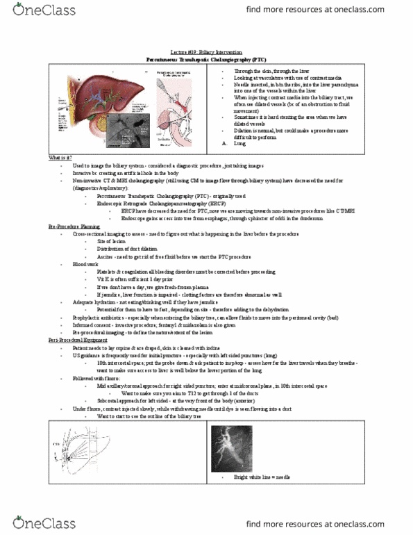
Produces a homogenous shadow beneath right dome of diaphragm, tapering to left. The organs and ducts participating in secretion (liver), storage (cystic duct and gall bladder) and delivery(hepatic and bile ducts) of bile into the duodenum. Cholangiopancreatography: radiographic examination of bile ducts and pancreatic ducts. Administration of contrast medium: direct injection into the duct through. Emphasizes difference between parenchyma and poorly enhancing lesions. Scans of different time intervals after contrast administration allow visualisation of different phases of opacification, enabling distinction of lesions such has hemangiomas and neoplasms. Non enhanced ct: calcifications, fat in tumours and fat-stranding. Arterial phase: rapid rise in aortic enhancement and gradual hepatic enhancement. Portal venous phase: contrast diffuses from central blood compartment to extravascular liver compartment (increase in hepatic enhancement and decrease in aortic) Equilibrium (delayed) phase: aortic and hepatic enhancement gradually decline as contrast diffuses back into central vascular compartment and to muscle and fat compartments. Higher the concentration, lower the flow rate.