NURS 163 Chapter Notes - Chapter 2: Peritoneum, Yolk Sac, Merocrine


Study Guide 2: Tissues Answers:
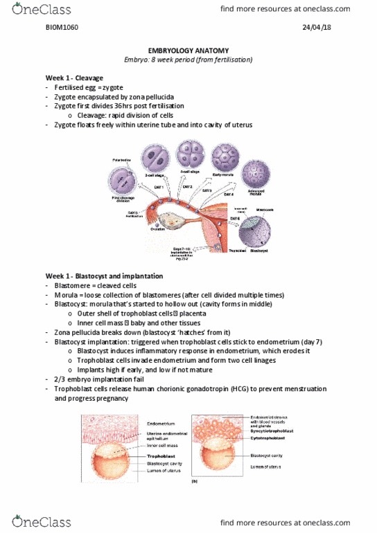
1. First stage of Pre-embryonic development: Cleavage
A. rapid mitotic divisions of a zygote
B. Cells divide smaller and smaller into many cells
C. Large surface area= large uptake of nutrients, oxygen
D. Blastomeres: building blocks for embryo
2.
A. Morula: >16 cluster of blastomere cells
a. 4-5 days after fertilization
b. Zona pelucida covering breaks down
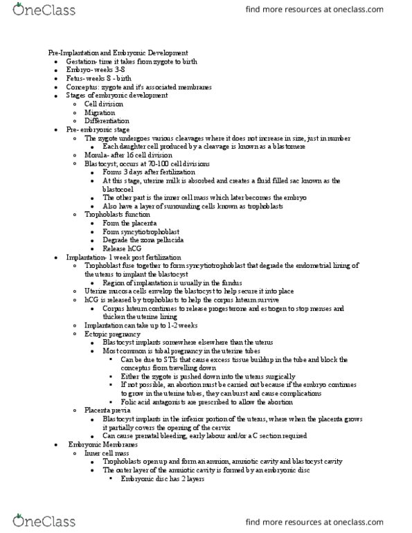
B. Blastocyst: hatches from morula no zona pelucida contains 2 cell types
3.
A. Trophoblasts: single outer layer of flattened cells on outer wall
a. w/ L section adhesion proteins
B. Inner cell mass: 20-30 round cell cluster on one side of cell
*See picture in Textbook
4. Trophoblast Function:
A. Protect inside cells
B. Eventually forms placenta
5. hCG role in Pregnancy? How is it useful?
A. Secreted from trophoblasts: prevents menstruation during pregnancy by
maintaining uterine lining
B. Still secretes progesterone & estrogen
C. Pregnancy tests: test for hCG levels in urine or blood
6. Bilaminar Embryonic Disc origination:
A. Inner cell mass becomes Bilaminar embryonic disc
B. Develops into two layers: epiblast & hypoblast w/ amniotic cavity in between
7. Three primary Germ Layers: (trilaminar disc)
1) Endoderm: develops from hypoblast
a. Forms: ET of GI, Respiratory, Urogenital systems & glands
2) Ectoderm: develops from epiblast
a. Forms: nervous system, skin epidermis
3) Mesoderm: develops from primitive streak
a. Forms: every other part of body
*See picture of these layers in Textbook
8. Four extraembryonic membranes & functions:
1) Amnion: encloses amniotic cavity (only broken by umbilical cord)
find more resources at oneclass.com
find more resources at oneclass.com

a. Bag of waters protects/supports embryo
2) Yolk Sac: under the hypoblast, (provides nutrients for bird fetus)
a. Forms: early blood cells, digestive structure, stem cells for
sperm/eggs
3) Allantois: out-pocketing of embryonic tissue at caudal of yolk sac
a. Base of umbilical cord, site of waste disposal, forms bladder
4) Chorion: outermost membrane
a. Encloses embryo, forms placenta
*See picture of these layers in Textbook
9. Tissue: group of similar cells, specialized for specific function
10. 4 Tissue Types and Function:
1) Epithelial: covering/lining
2) Connective: binding, support, insulation
3) Muscle: contraction
4) Nervous: information conduction
11. Three features to anchor cells, stabilize tissue:
1) Glycoproteins: cell surface
2) Basement Membranes: supports, anchors cells in place
3) Intercellular junctions: between adjacent cells
12. Five Intercellular Junctions: Structure and Function:
1) Tight Junctions: fusing transmembrane proteins of adjacent cells
a. Anchor along cell wall on all sides of PM
2) Adherens Junctions: dense protein (plaque) attaches membrane proteins &
cytoskeletal proteins
a. Cadherins: (CAM: cell adhesion molecule) transmembrane
glycoproteins, attach to plaque, attaches to cadherins of other cell
3) Desmosomes: (similar to adherens junctions)
a. Filaments on plaque attach to other desmosomes
b. Very strong, stable attachment for high stress areas
4) Hemidesmosomes: 2 layers of plaque
a. Transmembrane integrin proteins attach to laminas on other cell
b. Filaments attach to plaque
c. Anchor cells to basement membrane!
5) Gap Junctions: adjacent, close cells with fused membrane proteins
a. Connexons: hollow tubes formed by used proteins
b. Used for intercellular chemical transfer
*See picture of these layers in Textbook
13. Two Epithelial Tissue Categories:
1) Covering/Lining ET:
2) Glandular ET:
a. ET cells specialized to produce/secrete secretion: mucus
find more resources at oneclass.com
find more resources at oneclass.com

b. Can be endocrine or exocrine
14. Special ET Characteristics:
A. High degree cellularity: no matrix
B. High Regenerative capability: rapid mitosis= rapid regeneration
C. Specialized lateral contacts: intercellular junctions
D. Polarity: apical and basal surfaces
E. Avascular: BV in CT supply the ET through diffusion
F. Basement Membrane: anchors/supports ET
15. Lateral contacts in ET:
A. Tight Junctions
B. Desmosomes
C. Hemidesmosomes: anchor ET to basement membrane
16. ET Polarity: top and bottom surfaces different
A. Apical: free surface exposed to cavity or exterior (lumen)
B. Basal: not exposed, rests on basement membrane
17. Basement Membrane Function:
A. Anchors, supports ET
B. Selective permeability of diffusion
18-19. Two layers of Basement membrane
1) Basal Lamina: superficial layer
a. Thin layer secreted by ET cells
b. Selective filter
2) Reticular Lamina: deep layer
a. Collagen fibers from underlying CT fibroblasts
20. Covering and Lining ET Functions:
A. Protection
B. Control Permeability: absorption/secretion
C. Surface Transport: via cilia, spreads mucus
D. Sensory function
21. ET Cell Shape and Cell Layers:
A. Cell Layers
1. Simple
2. Stratified
3. Pseudostratified
B. Cell Shape
1. Squamous: thin, flattened
2. Cuboidal: cube-shaped
3. Columnar: long, narrow
*Transitional: cells change shape based on stretch
find more resources at oneclass.com
find more resources at oneclass.com
Document Summary
*transitional: cells change shape based on stretch: simple vs. *pathway for bv/nerves/lymph vessels: superficial fascia (hypodermis), areolar & adipose, deep fascia, multiple layers, dense ct: strong, resists stretch, anchors visceral organs, subserous fascia, between deep fascia and serous membranes. Serous membrane: lines cavities not opening to exterior: mucous membrane: lines cavities opening to exterior, cutaneous membrane: skin. Cadherins: transmembrane glycoproteins forming the adherens junctions by attaching to an adjacent cell"s plaque. Adhesion belt: areas of extensive adherens junctions, present in et. Integrin: in the basement membrane, attach to laminins on outsides of cells. Laminins: glycoproteins on basement membrane aiding in hemidesmosome adhesion. Connexons: fused transmembrane proteins in pm forming cylinders connecting adjacent cells in gap junctions. Peritoneum: serous membrane lining cavity and organs of abdominal/pelvic. Endothelium: simple squamous et lining walls of heart, bv, lymphatic vessels. Apical surface: superficial, free surface in et exposed to exterior/cavity. Basal surface: deep surface of et lining basement membrane.