BIOL 142 Chapter Notes - Chapter 9-10: Elastin, Loose Connective Tissue, Endocardium

9 Jul 2018
School
Department
Course
Professor
Document Summary
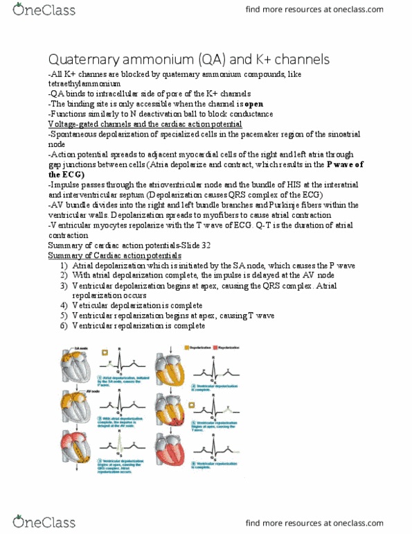
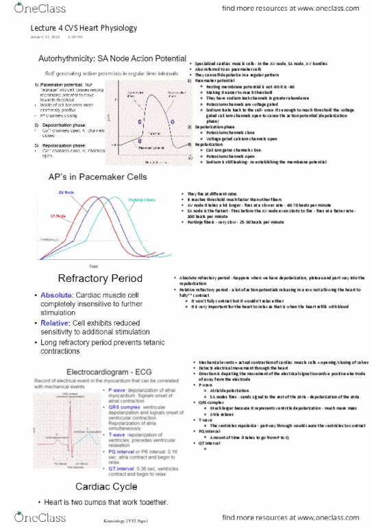
The av bundle splits into two pathways called this. Subendocardial fibers (purkinje fibers) each bundle branch gives rise to these which branch extensively and deliver action potentials the contractile myocytes composing the free (outer) walls of the ventricles. Electrocardiograph the machine used to produce a picture of the small voltage changes. Electrocardiogram the picture of the voltage changes across the body caused by cardiac action potentials. P wave wave created as cardiac action potentials depolarize the cells of the atria. Qrs complex/segment caused as cardiac action potentials depolarize the cells of the ventricle. T wave wave caused as the cells of the ventricles go through repolarization phase of the cardiac action potential. P-q segment (interval) the time from the end of the p wave, to the beginning of the qrs complex equal to the time between atrial depolarization and ventricular depolarization. S-t segment elevation common hallmark indicating cell damage and that a heart attack has occured.