PSYCH 1 Chapter Notes - Chapter 8: Retina, Scotopic Vision, Visual Acuity

Document Summary
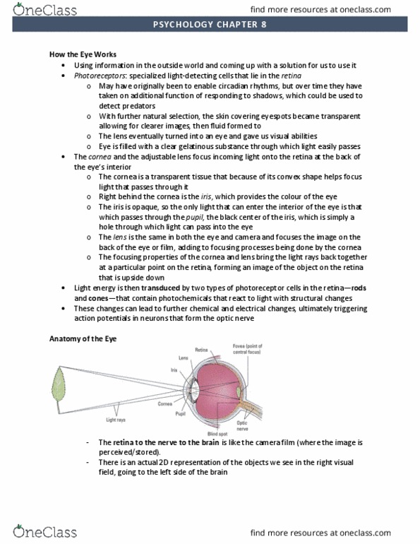
Brain scientists have estimated that somewhere between 25-40% of the human brain is devoted exclusively or primarily to the analysis of input from the eyes. In many species of multicellular animals, specialized light-detecting cells called photoreceptors evolved and became connected to the animal"s nervous system. Cross-species comparisons, based on homologies, suggest that the modern vertebrate eye came about through the following evolutionary steps: In some early ancestry to vertebrate animals, photoreceptors became concentrated into groups, forming light-detecting organs, or eye spots, just under the skin. Over time, these structures responded to shadows. Photoreceptors lie in the retina, a membrane lining the rear interior of the eyeball. The eyeball is filled with a clear gelatinous substance, called the vitreous humor, through which light easily passes. The cornea is a transparent tissue that, because of its convex (outward) curvature, helps focus the light that passes through it. Immediately behind the cornea is the pigmented, iris, which provides the color of the eye.