BIOL-242 Chapter Notes - Chapter 25: Renal Pelvis, Renal Artery, Renal Vein

9 Jun 2020
School
Department
Course
Professor
Document Summary
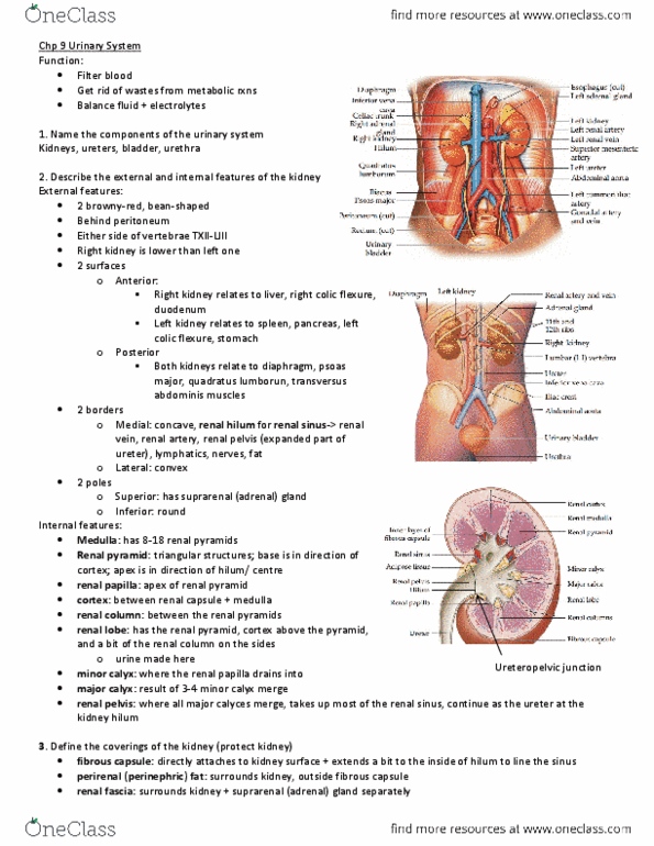
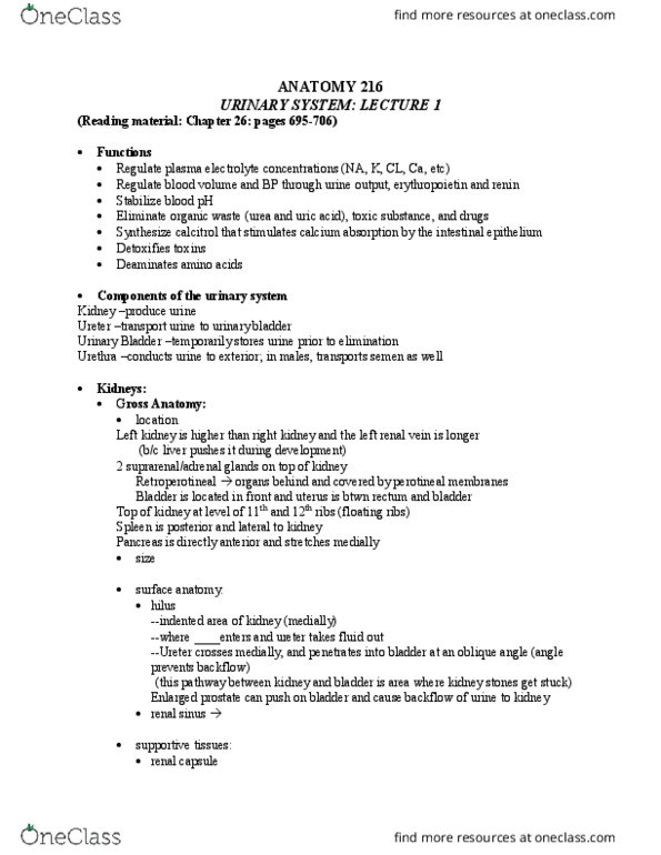
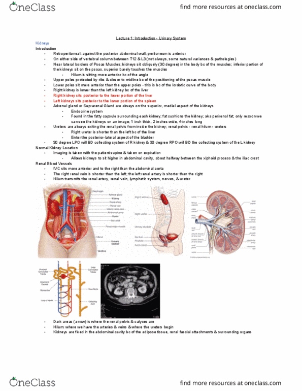
Describe the path that the ureter takes from the kidney to the bladder. Ureter is a continuation of the renal pelvis, originating from anterior to the renal vein and the renal artery. Crosses over the psoas muscle and iliac artery. Then joins the posterior wall of the urinary bladder. The right kidney sits below the __________ while the left kidney sits below the. The right kidney is tucked underneath the liver and the left kidney is inferior to the spleen and more medial. Kidney stones are condensed calcium crystals that form within the urinary system. A ct image can show detailed pictures of bone because it is composed of a dense organic matrix, which is primarily calcium salts. Kidney stones would look like round, white spheres on the ct scan, because kidney stones are composed of dense calcium crystals. Highly dense bone appears white on a ct image, whereas soft tissue has a more gray coloring.