PHIL 2015 Chapter Notes - Chapter 9.4: Epidermal Growth Factor Receptor, Submandibular Gland, Erbb

23 May 2020
School
Department
Course
Professor
Document Summary
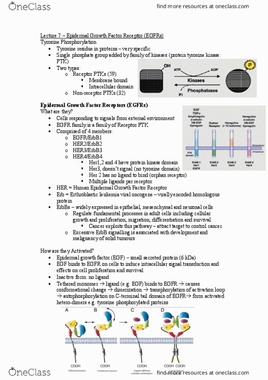
Stanley cohen isolated egf from an extract of the sub-maxillary gland and subsequently identified the egf receptor. Describe how the experiments that produced the data shown in figures 1 and 2 below enabled his group to start elucidating the first few events occurring soon after egf binds to the receptor. Figure 1 shows that in the presence of egf (upper curve) there was more phosphate incorporated from atp into cell membranes than in the absence of egf (lower curve). Figure 2 showed that the phosphate that was incorporated was actually binding to tyrosine and forming phosphotyrosine, showing that the receptor itself was being phosphorylated on its tyrosine residues. Knowing the molecular mechanisms underlying the activation of the egf (erbb) receptors, use the figure below and discuss how the receptors are kept inactive in the absence of ligands and how egf leads to their activation.