GASB05H3 Chapter Notes - Chapter 3: Temporal Lobe, Developmental Disorder, Extrastriate Cortex


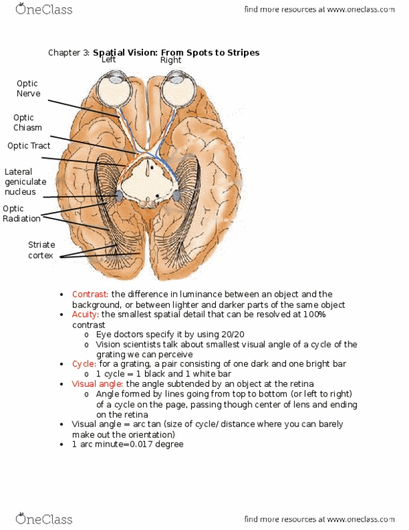
Chapter 3: Spatial Vision: From Spots to Stripes
- Acuity: the smallest spatial detail that can be resolved at 100% contrast
- Cycle: for a grating, a pair consisting of one dark bar and one bright bar
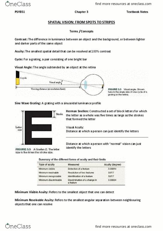
- Visual Angle: the angle subtended by an object at the retina
- Sine Wave Grating: A grating with a sinusoidal luminance profile as
shown in Figure 3.4a (on the right)
- Herman Snellen defines visual acuity as follows:
Types of Visual Acuity
- Minimum Visual Acuity
oMinimum visible acuity refers to the smallest object that one can detect
oBenito Daca de Valdez (1591 – 1634): measure the distance at which a roq of
mustard seeds could no longer be counted and early astronomers like Robert
Hooke (1635 – 1703) were interested in the size of stars that could be detected
and their relation to retinal anatomy
In this context the minimum visible acuity refers to the smallest target that
can be detected
Under ideal conditions, humans can detect a long, dark wire
against a very bright background when they subtend an angle of
just 0.5 arc second
It is widely accepted that the minimum visible acuity is so small for
two reasons
1. The optics of the eye spread the image of the thin line, making
it much wider on the retina
2. The fuzzy retinal image of the line costs a shadow that
reduces the light on a row of cones to a level is just detectably
less than the light on the row of cones on either side
oThe minimum visual acuity is actually limited by our ability to discriminate the
intensity of the target relative to its background
- Minimum Resolvable Acuity
oMinimum resolvable acuity refers to the smallest angular separation between
neighboring objects that one can resolve
oIt represents one of the fundamental limits of spatial vision: It is the finest high –
contrast detail that can be resolved. In foveal vision the limit is determined
primarily by the spacing of photoreceptors in the retinas
find more resources at oneclass.com
find more resources at oneclass.com

- Minimum Recognizable Acuity
oRefers to the angular size of the smallest feature that one can recognize or
identify
- Minimum Discriminable Acuity
oRefers to the angular size of the smallest change in a feature (change in size,
position, or orientation) that one can discriminate
- Acuity for Low – Contrast Stripes
oSpatial Frequency: the number of grating cycle (dark and bright bars) in a given
unit of space
oCycles per Degree: the number of grating cycles per degree of visual angle
oContrast sensitivity function: a function describing how the sensitivity to contrast
(defined as the reciprocal of the contrast threshold) depends on the spatial
frequency (size) of the stimulus
oContrast Threshold: the smallest amount of contrast required to detect a pattern
- Retinal Ganglion Cells and Stripes
oWhen the spatial frequency of grating is too low the ganglion cell responds
weakly because part of the fat, bright bar of the grating lands in the inhibitory
surround, damping the cell’s response
oWhen the spatial frequency is too high, the ganglion cell responds weakly
because both dark and bright stripes fall within the receptive – field center,
washing out the response
oWhen the frequency is just right with a bright bar filling the center and dark bars
filling the surround the cell responds vigorously
Therefore the retinal ganglion cells are tuned to spatial frequency; each
cell responds best to a specific spatial frequency that matches its
receptive field size, and it responds less to both higher and lower spatial
frequencies
oPhase: the relative position of a grating
- The Lateral Geniculate Nucleus
oThe axons of retinal ganglion cells synapse in the two lateral geniculate nuclei
(LGNs): One in each cerebral hemisphere
They act as relay station on the way from the retina to the cortex
The LGN of primates is a six layered structure
The neurons in the bottom two layers are physically larger than
those in the top four layers;
find more resources at oneclass.com
find more resources at oneclass.com

This is why the bottom two are called magnocellular layers and
the top four are called parvocellular layers
oThe magnocellular layers receive inputs from the M
ganglion cells in the retina
They respond to large fast moving objects
oThe parvocellular layers receive inputs from the P ganglion
cells
They are responsible to process the details of
stationary targets
Koniocellular cell: a neuron located between the magnocellular
and parvocellular layers of the lateral geniculate nucleus
oFrom the bottom to top layers 1,4 and 6 of the right LGN
receives input from the left (contralateral) eye
oWhile layers 2,3, and 5 get their input from the right
(ipsilateral) eye
Each LGN layer contains a highly organized map of a complete half of the
visual field
Topographical Mapping: provides us with a neural basis for knowing
where things are in space
- The Striate Cortex
oPrimary visual cortex (V1), area 17 or striate cortex: the area of the cerebral
cortex of the brain that receives direct inputs from the lateral geniculate nucleus
as well as feedback from other brain areas
oA major and complex transformation of visual information takes place in the
striate cortex
Cortical magnification: the amount of cortical area (usually specified in
millimeters) devoted to a specific region
oVisual crowding: the deleterious effect of clutter on peripheral object recognize
- Receptive Fields in Striate Cortex
oOrientation tuning: the tendency of neuron in striate cortex to respond optimally
to certain orientations and less to other
- Other Receptive – Field Properties
find more resources at oneclass.com
find more resources at oneclass.com
Document Summary
Chapter 3: spatial vision: from spots to stripes. Acuity: the smallest spatial detail that can be resolved at 100% contrast. Cycle: for a grating, a pair consisting of one dark bar and one bright bar. Visual angle: the angle subtended by an object at the retina. Sine wave grating: a grating with a sinusoidal luminance profile as shown in figure 3. 4a (on the right) Herman snellen defines visual acuity as follows: Hooke (1635 1703) were interested in the size of stars that could be detected and their relation to retinal anatomy. In this context the minimum visible acuity refers to the smallest target that can be detected. Under ideal conditions, humans can detect a long, dark wire against a very bright background when they subtend an angle of just 0. 5 arc second. In foveal vision the limit is determined primarily by the spacing of photoreceptors in the retinas.