PSYCH261 Chapter Notes - Chapter 6: Membrane Potential, Ciliary Muscle, Vitreous Body

1 May 2018
School
Department
Course
Professor

Vision
Mrs.R- cannot recognize objects, can name them once she picks them up be not from just looking.
Cannot recognize faces. Had stroke. Damage to ventral stream
Sensory receptors- specialized neuron that detects a particular category of physical events. Event
changes membrane potential which causes action potential
Sensory transduction- process of event cause AP
Receptor potentials- slow, graded electrical potential caused by change in physical stimuli
The Stimulus
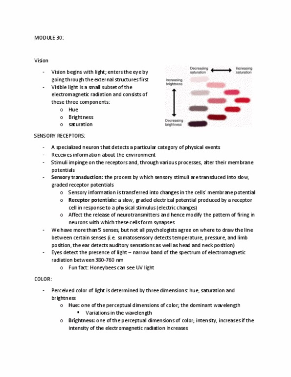
Colour- based on three dimensions; hue, saturation, and brightness.
Hue- determined by wavelengths, we can only see some wavelength aka hues
Brightness- varies in intensity, increase in electromagnetic radiation is increase in brightness
Saturation- purity of light that is being perceived, if all radiation is one wavelength it is more pure, if all
visible wavelengths are present then it is very saturated
Anatomy
Orbits-bony pockets in the front of the skull
Sclera- tough, white outer coat of eye, where muscle attach, does not allow light through
Conjunctiva- mucous membrane lining eyelid and attaching to eye, prevent contacts from going behind
eye
Vergence movements- keeps eye fix on target, going crossed eyed, eyes moving together to keep image
equal in both retinas
Saccadic movements- jerky movements as eyes scan scene
Pursuit movement- slow eye scan, must focus on object as it slowly moves, aka finger moving in front of
you
Cornea- outer layer in front of eye, allows light in
Pupil- regulated amount of light entering eye, opening in the iris, pigmented ring of muscle stimulated
by cornea
Lens- behind iris, made up of many transparent layers, ciliary muscles alter its shape.
Accommodation- extension and flexion of ciliary muscle, focus images on retina
Vitreous humor- liquid of the eye, in between lens and retina
Retina- interior lining of back of the eye, has receptor cells in it, neural tissue and photo receptors
Rods- rod shaped, sensitive to light at low intensities, one type of photo receptor
find more resources at oneclass.com
find more resources at oneclass.com

Cones- cone shaped, sensitive to one of three wavelengths of light that humans can see, give us info
about small features, important in daytime vision
Fovea- area in center of retina, mediates most acute vision, contains only cones,
Optic disk- exit point on retina for ganglion cells that form optic nerves, images cannot be projected on
this area causing a blind spot
Bipolar cells-photoreceptors form synapses with them. Connect to shallowest and deepest layers of
retina. Middle layer retinal neurons that pass information from photoreceptors to ganglion cells
Ganglion cell- connects to bipolar cell to receive info. Axons ravel through optic nerve, carry into the rest
of the brain
Horizontal cells- in retina. Connects photoreceptors and outer processes of bipolar cells
Amacrine cells- in retina, connects ganglion and inner bipolar cells.
Photoreceptors
Lamella- thin plates of membrane, containing photopigments, found in rods and cones
Photopigments- special molecules in lamella, rods contain about 10 million, responsible for transduction
of visual information. Comes from Vitamin A. made up of opsin and retinal
Opsin- a protein, several types including rod opsin found in the photopigment rhodopsin
Retinal- a lipid, synthesized from vitamin?[
Rhodopsin- before meeting light it is pink. When exposed to light it breaks into rod opsin and retinal,
olour hages to a yellow, therefore we say light leahes photopigets. The splittig reates a
receptor potential
Action potential- not produced by photoreceptors or bipolar cells, they release a neurotransmitter
instead. Light striking a photoreceptor produces a hyperpolarization, so the photoreceptor releases less
neurotransmitter. Because the neurotransmitter normally hyperpolarizes the membrane of the bipolar
cell, the reduction causes a depolarization. This depolarization causes the bipolar cell to release more
neurotransmitter, which excites the ganglion cell.
Connection Between Eye and Brain
Dorsal lateral geniculate nucleus- LGN. axons from retinal ganglion descend to connect here. Part of
thalamus. Made up of six layers, each layer only gets info from one eye. Projects info onto primary
visual cortex
Magnocellular layers-inner two layers of LGN. Transmits perception of form, movement, depth, and
brightness to primary visual cortex
Parvocellular layers- other four layers of LGN, transmits colour and fine detail info
Koniocellular sublayers- found ventral to magno and parvo. Transmit short wavelength cone info to
primary visual cortex
find more resources at oneclass.com
find more resources at oneclass.com

Calcarine fissure- horizontal fissure on posterior cerebral cortex. Contains optic radiations (where axons
from LGN travel to primary neuron cortex). Location or primary visual cortex
Striate cortex- another name for primary visual cortex, named this because it contains dark-staining
layer of cells
Optic chiasm- where optic nerves join together in an x-shape, ganglion cells with inner retina info cross
over to the other side and their axons descend to LGN. Outer retinal info does not cross over
Coding of Light and Dark
Receptive field- area of visual field where stimuli will produce an alteration in firing rate of particular
neuron. Location depend on specific neuron location. Fovea receptive fields will be at a fixed point,
peripheral neurons have fields off to one side
Ratios- more ganglion to cones in fovea than in peripheral. Why we have better vision in fovea
Hartline(1938)- discovered three types of retina ganglion cells in frogs. ON cells respond to burst on
retina, OFF cells when there is no light, ON/OFF responds when light is first turn off and turned off
Schiller(1992)- ganglion fire normally at very low rate, ON cells increase rate, OFF cells decrease rate
Coding of Colour
Thomas Young (1802)-eye contain three different receptors, one for each hue. All colors on visible
spectrum can be made from colour mixing(not the same as paint mixing). Addition of two or more light
sources
Ewald Hering(1905)- primary colour thought to be red, blue, green, and yellow, do not seem to blend.
Trichromatic coding- young was right. Each receptor absorbs different wavelengths. Characteristics are
determined by different opsins. Red cones evolved into blue cones, red than mutated to green
find more resources at oneclass.com
find more resources at oneclass.com
Document Summary
Mrs. r- cannot recognize objects, can name them once she picks them up be not from just looking. Sensory receptors- specialized neuron that detects a particular category of physical events. Event changes membrane potential which causes action potential. Receptor potentials- slow, graded electrical potential caused by change in physical stimuli. Colour- based on three dimensions; hue, saturation, and brightness. Hue- determined by wavelengths, we can only see some wavelength aka hues. Brightness- varies in intensity, increase in electromagnetic radiation is increase in brightness. Saturation- purity of light that is being perceived, if all radiation is one wavelength it is more pure, if all visible wavelengths are present then it is very saturated. Orbits-bony pockets in the front of the skull. Sclera- tough, white outer coat of eye, where muscle attach, does not allow light through. Conjunctiva- mucous membrane lining eyelid and attaching to eye, prevent contacts from going behind eye.