PSYCH261 Chapter Notes - Chapter 8: Gamma Motor Neuron, Golgi Tendon Organ, Tendon Reflex

1 May 2018
School
Department
Course
Professor

Control of Movement
Skeletal muscles
Skeletal muscles- striated muscle attached to bone. Connected to bones via tendons
Antigravity muscles- muscles used to stand up
Extrafusal muscles- muscle fiber that is responsible for force exerted when contracting muscle
Alpha motor neurons- synapse to extrafusal muscles. Activate contraction
Intrafusal muscles- specialized sensory organs. Serve two axons, motor and sensory. Also called muscle
spindles. Stretch receptor. Parallel to extra.
Gamma motor neuron- causes intrafusal to contract
Neuromuscular junction- synapse between terminal button and muscle fiber (motor endplate)
Endplate potential- postsynaptic potential needed to release Ach
Golgi tendon organ- between muscle and tendon. Sensitive to stretch
Reflexes
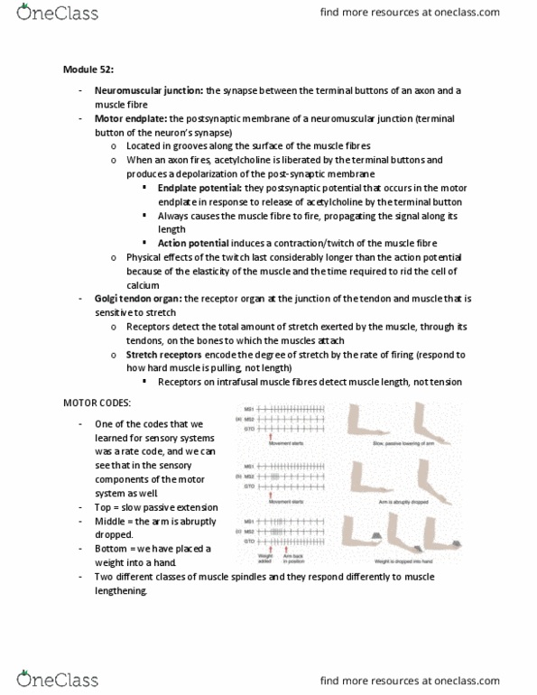
Mono synaptic stretch reflexes- patellar tendon reflex. Only includes one synapse, no brain involvement
Gamma motor neurons- when activated they make muscle spindles smaller and therefore more
sensitive. More precious control of movement. Establishes rate of fire
Polysynaptic reflexes- more than one synpase
Interneurons- not afferent or motor, inbetween them, found in spine. Used to slow down reflex
Movement Controlled by the Brain
Somatotropin organization- way of mapping brain based on what part of motor cortex controls body
find more resources at oneclass.com
find more resources at oneclass.com

Supplementary motor area - beside to primary motor cortex, medial surface of brain. Receive info from
parietal and temporal lobes and send to primary
Premotor cortex- beside to primary motor cortex, lateral surface of brain. Receive info from parietal and
temporal lobes and send to primary
Lateral group- corticospinal tract, corticobulbar tract and the rubrospinal tract. Control of independent
limb movement
Ventromedial group- contains vestibulospinal tract, the tectospinal tract, the reticulospinal
tract, and the ventral corticospinal tract. Control autonomic movements: gross movements of trunk,
posture and locomotion.
Corticospinal tract- contain axons from cortical neurons to grey matter in spine. Axons leave motor
cortex to enter the peduncles in the medualla
Pyramid tracts- what axons become when leaving peduncles. Found on ventral border of mendulla
Lateral corticospinal tract- axons that come from motor cortex to contralateral ventral grey matter.
Controls movement of distal limbs
Ventral corticospinal tract- axons from motor cortex to ipsilateral ventral grey matter. Controls upper
legs and trunk
Corticobulbar tract- projects into medulla. Terminates in the 5, 7, 9, 10, 11 and 12th cranial nerve.
Control face, neck, tongue and eyes.
Rubrospinal tract- originates in red nucleus of midbrain, terminates on spinal cord. Controls
independent movements of forearms and hands
Vestibulospinal tract-go from vestibular nuclei to gray matter in spine. Control posture in response to
vestibular system
Tectospinal tract- go from tectum to gray matter. Controls head and trunk movements to match with
eyes
Reticulospinal tract- go from reticular formation to gray matter in spine
find more resources at oneclass.com
find more resources at oneclass.com
Document Summary
Extrafusal muscles- muscle fiber that is responsible for force exerted when contracting muscle. Neuromuscular junction- synapse between terminal button and muscle fiber (motor endplate) Endplate potential- postsynaptic potential needed to release ach. Gamma motor neurons- when activated they make muscle spindles smaller and therefore more sensitive. Interneurons- not afferent or motor, inbetween them, found in spine. Somatotropin organization- way of mapping brain based on what part of motor cortex controls body. Supplementary motor area - beside to primary motor cortex, medial surface of brain. Premotor cortex- beside to primary motor cortex, lateral surface of brain. Receive info from parietal and temporal lobes and send to primary. Lateral group- corticospinal tract, corticobulbar tract and the rubrospinal tract. Ventromedial group- contains vestibulospinal tract, the tectospinal tract, the reticulospinal tract, and the ventral corticospinal tract. Control autonomic movements: gross movements of trunk, posture and locomotion. Corticospinal tract- contain axons from cortical neurons to grey matter in spine.