BPK 142 Chapter Notes - Chapter 6.0: Bronchitis, Tidal Volume, Chronic Obstructive Pulmonary Disease

22 Aug 2018
School
Department
Course
Professor

1
I.
Anatomy of the Respiratory System
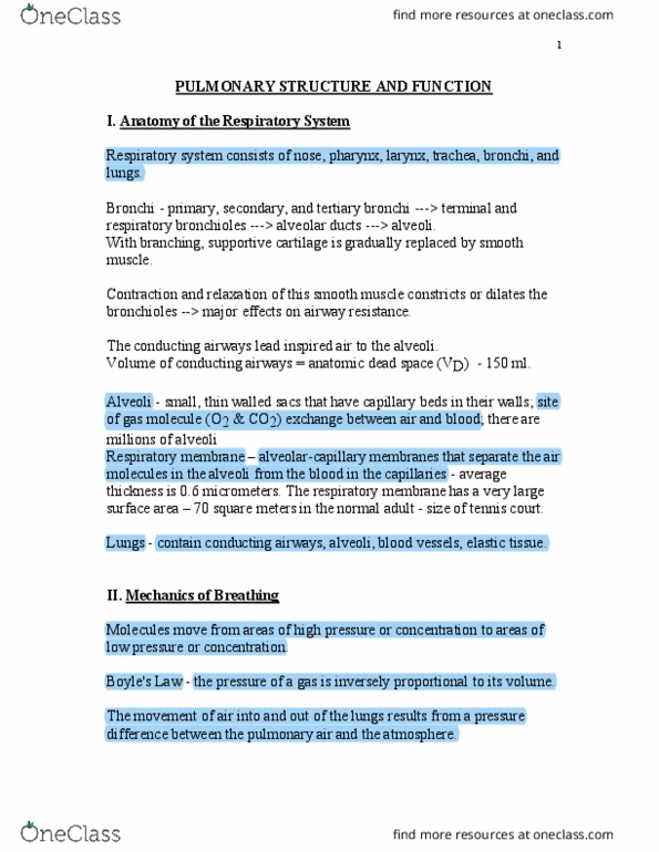
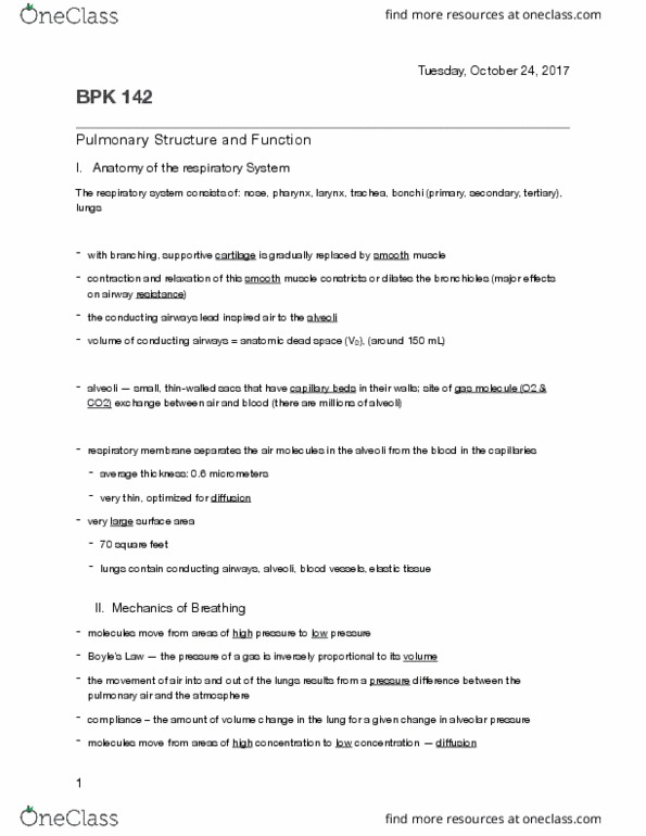
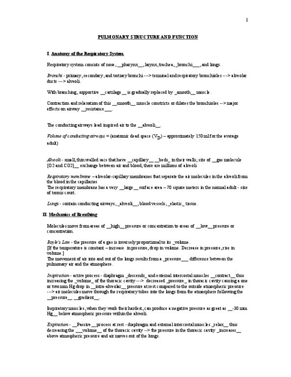
Respiratory system consists of nose, pharynx, larynx, trachea, bronchi, and lungs.
Bronchi - primary, secondary, and tertiary bronchi ---> terminal and respiratory
bronchioles ---> alveolar ducts ---> alveoli.
With branching, supportive cartilage is gradually replaced by smooth muscle.
Contraction and relaxation of this smooth muscle constricts or dilates the bronchioles
--> major effects on airway resistance.
The conducting airways lead inspired air to the alveoli.
Volume of conducting airways = anatomic dead space (VD) - 150 ml.
Alveoli - small, thin walled sacs that have capillary beds in their walls; site of gas
molecule (O2 & CO2) exchange between air and blood; there are millions of alveoli
Respiratory membrane – alveolar-capillary membranes that separate the air molecules
in the alveoli from the blood in the capillaries - average thickness is 0.6 micrometers.
The respiratory membrane has a very large surface area – 70 square meters in the
normal adult - size of tennis court.
Lungs - contain conducting airways, alveoli, blood vessels, elastic tissue.
II.
Mechanics of Breathing
Molecules move from areas of high pressure or concentration to areas of low
pressure or concentration.
Boyle's Law - the pressure of a gas is inversely proportional to its volume.
The movement of air into and out of the lungs results from a pressure difference between
the pulmonary air and the atmosphere.

2
Inspiration - active process - diaphragm descends and external intercostal muscles
contract thus increasing the volume of the thoracic cavity ---> decreased pressure in
thoracic cavity causing a one or two mm Hg drop in intra-alveolar pressure at rest
compared to the outside atmospheric pressure
---> air molecules move through the respiratory tubes into the lungs from the
atmosphere following the pressure gradient.
Inspiratory muscles, when they work their hardest, can produce a negative pressure as
great as -30 mm Hg below atmospheric pressure within the alveoli.
Expiration - passive process at rest. Secondary muscles, such as abdominal
muscles become involved in exercise.
Forced expiration can produce intra-alveolar pressure as great as +50 mm Hg above
atmospheric pressure.
During exercise, mouth breathing tends to replace nasal breathing - less resistance
to airflow.
Air that enters the respiratory passages via either the nose or the mouth is quickly
saturated with water vapor and warmed to body temperature, 37 degrees centigrade,
even under conditions when very cold air is inspired.
Compliance – the amount of volume change in the lung for a given change in alveolar
pressure.
III.
Lung Volumes
Refer to Lab Manual, pages 17-4 and 17-5, for definitions of various lung volumes
and capacities.
Normal values at rest:
Minute ventilation (VE ) - 6 - 8 liters/min.
Tidal volume (VT) - 500 ml per inspiration or expiration
Breathing frequency (FR) - 12 - 16 breaths per minute
Expiratory reserve volume (ERV) – approximately 25% of vital capacity (VC)
Document Summary
Respiratory system consists of nose, pharynx, larynx, trachea, bronchi, and lungs. Bronchi - primary, secondary, and tertiary bronchi ---> terminal and respiratory bronchioles ---> alveolar ducts ---> alveoli. With branching, supportive cartilage is gradually replaced by smooth muscle. Contraction and relaxation of this smooth muscle constricts or dilates the bronchioles. The conducting airways lead inspired air to the alveoli. Volume of conducting airways = anatomic dead space (vd) - 150 ml. Alveoli - small, thin walled sacs that have capillary beds in their walls; site of gas molecule (o2 & co2) exchange between air and blood; there are millions of alveoli. Respiratory membrane alveolar-capillary membranes that separate the air molecules in the alveoli from the blood in the capillaries - average thickness is 0. 6 micrometers. The respiratory membrane has a very large surface area 70 square meters in the normal adult - size of tennis court. Lungs - contain conducting airways, alveoli, blood vessels, elastic tissue.