NSE 13A/B Chapter Notes - Chapter 22: Non-Alcoholic Fatty Liver Disease, Esophageal Varices, Abdominal Wall

8 May 2015
School
Department
Course
Professor
Document Summary
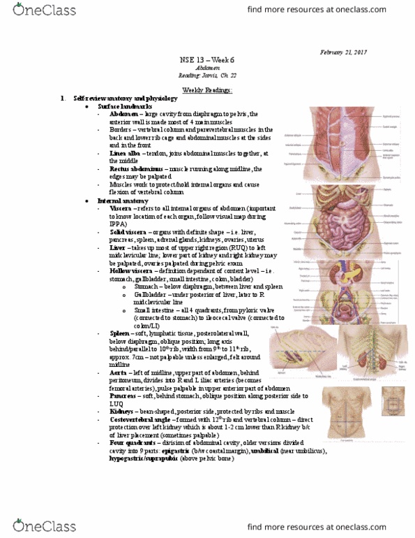
Chapter 22: abdomen: these are joined at the midline by a tendinous seam the linea alba. The abdomen is a large oval cavity extending from diaphragm down to top of pelvis. Four layers of large, at muscles form the ventral abdominal wall. The rectus abdominis forms a strip extending the length of the midline and its edge is often. Muscles protect and hold organs in place and they ex the vertebral column. All internal organs are called the viscera. Shape of hollow viscera (stomach, gallbladder, small intestine, colon, and bladder) depends on the contents: usually not palpable the umbilicus. The abdominal wall is divided into four quadrants by a vertical and horizontal line bisecting. Left spermatic cord costal margin: in healthy full-term newborns, lower edge may be palpated 0. 5 to 2. 5cm below right diarrhea high turnover for water and electrolytes. Abdominal wall is less muscular so organ may be easier to palpate.