MEDRADSC 2Z03 Chapter Notes - Chapter 6: Angiography, Brain Injury, Ct Pulmonary Angiogram

27 Jun 2016
School
Department
Course
Professor
Document Summary
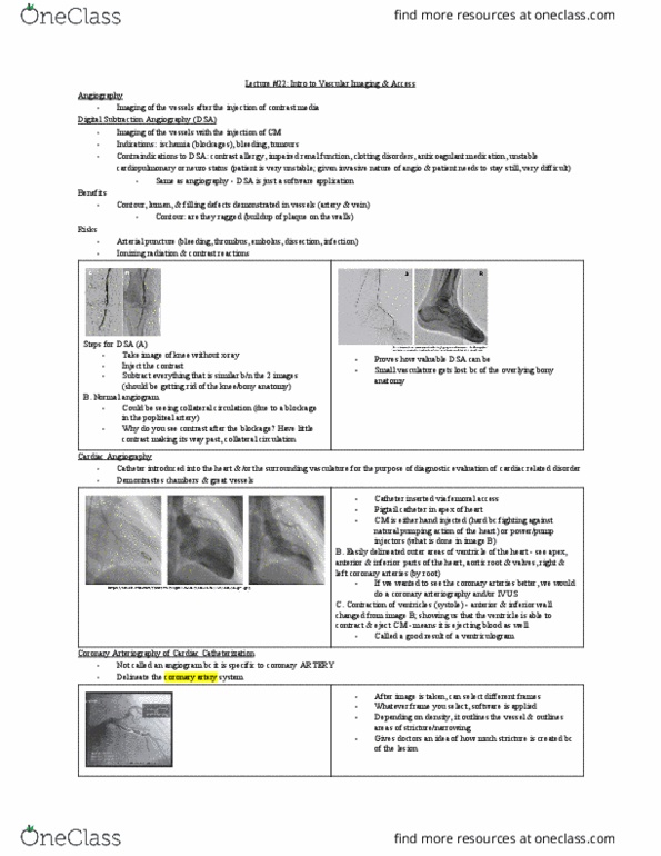
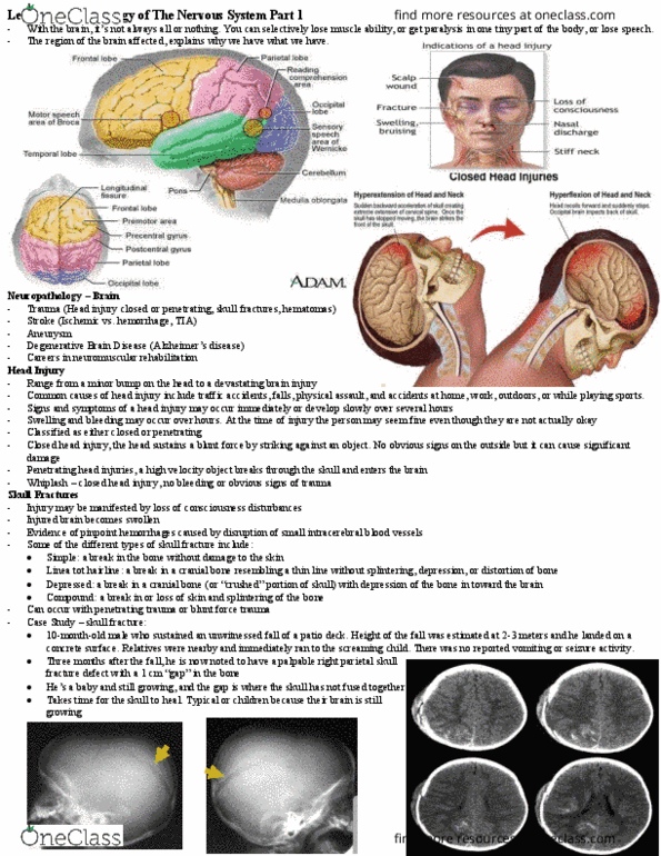
Cranial ct is an accurate test to delineate brain structures and can identify brain matter, arteries, veins, cerebrospinal fluid filled ventricles and the bony architecture of the skull. A ct scan, also called a cat scan, of the head (see images 1 and 2) is usually prescribed to detect: bleeding or blood clot formation, brain injury and skull fractures in patients with head injuries, brain tumours. Head ct scan (axial views) showing image of the brain: Image 1: normal brain image 2: brain with heamorrage. Ct angiography (see image 3 below) uses an injection of iodine-based x-ray dye into a vein, in order to see the vessels more accurately. Ct angiography will assess the severity of any narrowing of the arteries. intracerebral. Image 3: head scan, ct angiogram (coronal view) A ct scan of the chest may be performed. To assess the chest and its organs, including respiratory and cardiovascular systems, as well as the.