MEDRADSC 2Z03 Chapter Notes - Chapter 7: Sagittal Plane, Coronal Plane, Gastrocnemius Muscle

27 Jun 2016
School
Department
Course
Professor
Document Summary
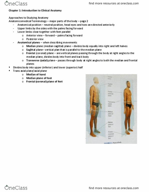
In the context of medical imaging, the body is divided into three orthogonal planes. Sagittal: this plane is parallel to the spinal column, and divides the body into left and right. The line passing directly through the spinal column is known as the medial sagittal plane. Axial (transverse): this plane divides the body into superior and inferior components. Coronal: this plane divides the body into anterior and posterior sections. There are certain international conventions to follow when acquiring and displaying ultrasound images. In sagittal (or longitudinal) images, the superior or head end of the patient is to the viewer"s left side. This can be seen below in figure 1 of the gastrocnemius muscle, where the superior and inferior aspects are marked. Before the scan, a full obstetrical history is taken. This date is entered into the ultrasound system, and estimated fetal age will be calculated and displayed as weeks and days, e. g. 18w 4d.