BIOLOGY 2B03 Chapter Notes - Chapter 7: Actomyosin Ring, Phalloidin, Ovarian Cancer

17 Jun 2018
School
Department
Course
Professor


BIOLOGY 2B03 - Module 7 Lecture I
Cytoskeleton is Highly Organized
● Cytoskeleton provides cell shape and structure
● Specialized cell structures is necessary in differentiated cells
● Cell shapes depend on the functions of different filaments
Cytoskeleton is Dynamic
● Dynamic to provide movement
○ ex) migration of cells and cell division
○ animation) ovarian cancer cell migration on glass; cell is expressing Actin-GFP
● Breast cancer cell undergoing cell division; expressing Tubulin:GFP
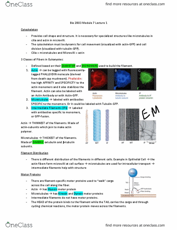
Cytoskeleton is a System of Filaments and Tubules
● Three classes of filaments in eukaryotic cells
○ Defined by diameter and the type of subunit used as building blocks for the filament
● Actin filaments are labeled with fluorescently-tagged phalloidin molecule
○ Phalloidin molecule: toxin derived from a mushroom (death cap)
○ Bind to actin monomer with high affinity and specificity
○ Stabilized the filament when bound
● Actin can be also labeled with antibody or with a protein fusion (Actin:GFP)
● Microtubules are labeled with antibodies specific to the subunits or protein fusion
(Tubulin:GFP)
● Intermediate Filaments (IFs): can be labeled as antibody specific to a monomeric subunit of
the filament of GTF-fusion
● They all form overlapping
Cytoplasmic Fibres are Polymers
● Each filament is constructed from smaller protein subunits to form a long polymer
● Actin filament: thinnest filament and are composed of monomeric actin subunits
● Microtubules: thickest filaments and are made up of dimeric subunits of alpha- and beta-
tubulin
● There are many different IFs; each is assembled from a different protein or set of proteins
Filament Distribution
● Three different ways of the filaments distributions in each cell type
● Epithelial cell: all three types of filaments are seen in unique locations
○ Actin: microvilli shape at the apical cell surface
○ IFs: provide structural support by spanning the cell
■ Made with lamin proteins form the nuclear lamina: provides structure and
shape to the nucleus
○ Microtubules: networks for intracellular
transport
Motor Proteins Track on Filament Highways
● There are filament-specific motor proteins
○ Track along actin filaments and microtubules
● No motor proteins identified for IFs
● Myosin Protein: move along actin filaments
● Kinesin and Dynein: track along microtubules
● The head domains bind to a cytoskeletal fiber (AFs or
MTs) and the tail domain attaches to a cargo
find more resources at oneclass.com
find more resources at oneclass.com

● ATP hydrolysis → provides energy
Actin-based Structures and Movement
● Highest density of actin is at the cell periphery
● Actin filaments underlying the cell membrane determine the shape and movement of the cell
surface
● Typical functions: the establishment of microvilli, the formation of contractile bundles that
form sarcomeres, formation of filopodia and lamellipodia
○ Sarcomeres: power muscle cell contraction
○ Filopodia and lamellipodia: needed for cell migration and the contractile ring that
directs cytokinesis
Actin Filaments are Assembled from Monomers
● Actin filament (F-actin, EM) are two-stranded helical polymers
● Total diameter 5-9nm
● Each polymer is built from actin monomer called G-actin
Actin Filaments are Polar
● Two ends look and behave differently from one another
● Actin filaments have been decorated with a portion of the actin-binding
protein, myosin
● Myosin: its head protein binds in just one orientation on the actin filament
○ Proteins point away from the actin filament
○ Able to define plus and minus end based on the rate of actin
polymerization
● The plus end: grows more quickly through addition of more actin subunits
and has barbed appearance
● The minus end: grows more slowly and may shrink, has a pointy appearance
G-actin Monomer
G-Actin: a single actin monomer
● Can be divided into four structural domains with a large cleft between
domains 2 and 4
○ The cleft forms an ATP-nucleotide binding site
● Each of the is polar → the microfilaments built up from these are also
polar
● The ATP-binding pocket is pointed to the minus-end
○ ATP-binding pocket of each monomer is not exposed within a
filament
○ Except for a pair of monomers right at the minus-end
F-actin: Actin Polymerization and Depolymerization
Filamentous Actin: is created through the polymerization of actin monomers
● Note: actin filament is not a static but dynamic structure
○ Constantly engaged in polymerization and depolymerization
○ Tendency to be more growth at the plus-end and more shrinkage at the minus-end
● ATP-binding regulates the growth and disassembly of the actin filaments
○ Enough [actin-ATP] in
cytosol → able to join
the plus end
Actin has an intrinsic ATPase activity
find more resources at oneclass.com
find more resources at oneclass.com
Document Summary
Specialized cell structures is necessary in differentiated cells. Cell shapes depend on the functions of different filaments. Ex) migration of cells and cell division. Animation) ovarian cancer cell migration on glass; cell is expressing actin-gfp. Breast cancer cell undergoing cell division; expressing tubulin:gfp. Cytoskeleton is a system of filaments and tubules. Three classes of filaments in eukaryotic cells. Defined by diameter and the type of subunit used as building blocks for the filament. Actin filaments are labeled with fluorescently-tagged phalloidin molecule. Phalloidin molecule: toxin derived from a mushroom (death cap) Bind to actin monomer with high affinity and specificity. Actin can be also labeled with antibody or with a protein fusion (actin:gfp) Microtubules are labeled with antibodies specific to the subunits or protein fusion (tubulin:gfp) Intermediate filaments (ifs): can be labeled as antibody specific to a monomeric subunit of the filament of gtf-fusion. Each filament is constructed from smaller protein subunits to form a long polymer.