BIOL 260 Study Guide - Midterm Guide: Endoplasmic Reticulum, Intercalated Disc, Skeletal Muscle

21 Sep 2016
School
Department
Course
Professor

Bio 260- test 3 notes round 2
2/24/16
- Three kinds of muscle
o Muscle tissue exerts force on other things (this is why we classify them together)
o Skeletal
o Cardiac
o Smooth
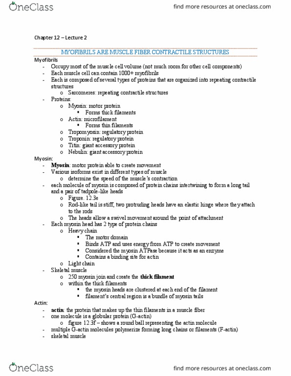
- All muscles depend on myosin and actin proteins
o Myosin
▪ Two sub units that are long polypeptide chains with heads on them
▪ The chains wrap around each other
▪ The heads have enzymatic activity which helps muscles pull
▪ The shafts interact with even more myosin shafts to make larger
structure
▪ The association of two myosin protein into a dimer is quaternary
structure
o Actin- the other critical protein
▪ Found in thin filaments
▪ Myosin heads want to interact with actin
• if they can they will crawl along the actin filament
• this is important because the myosin heads are found in thick
filaments where there are lots of them
• when they crawl along the actin, it pulls the thin filaments
• the actin thin filaments are anchored to other members of the
cytoskeleton, so the shape of the cell changes
• this is the method of muscle contraction in all three types of
muscle
- skeletal muscle
o all voluntary muscles we use to move- muscles you lift weights for
o skeletal muscle cells are very long and large in diameter
o the dark spots are nuclei, and each skeletal muscle cells can have multiple
o form by diffusion of multiple small starting cells
o once they form and mature, cannot go through mitosis
o striated- because myosin thick filaments and actin thin filaments are organized in
parallel fashion (with myosin being darker than actin)
o must do action potential before it can pull, to shorten the cell (contract)
o larger fiber like structures inside cell are myofiberals
o a bunch of myofiberals together can be called a skeletal muscle fiber
o myofiberals are surrounded by highly specified endoplasmic reticulum called a
sarcoplasmic reticulum.
▪ Specialized for calcium storage in muscle cells
▪ The sudden release of calcium from the sarcoplasmic reticulum is what
begins the contraction of the muscle
find more resources at oneclass.com
find more resources at oneclass.com

o the membrane of skeletal muscle cells does the action potentials
o Sarco and myo- related to muscles
- Cardiac muscle
o Only found in the heart- duh
o Look similar to skeletal muscle cells because they are striated, meaning the have
actin and myosin filaments in parallel repeating structures
o Much shorter than skeletal muscles- rather short actually
o Have much more complex shapes than skeletal muscle cells
o You will never see mitosis in cardiac cells
o But, you will see intercalated disc connecting the cells
▪ Intercalated disc- combined desmosomes and gap junction structures
▪ Desmosomes makes them mechanically tight (connecting cytoskeleton to
cytoskeleton) while the gap junction gives electric connectivity
o Cardiac muscle does not need neurons to stay on because of pacemaker cells
▪ Pacemaker cells depolarize themselves enough to do an action potential
which keeps the whole heart beating
- Smooth muscle
o Called smooth because it is not striated
o Very small
o Have one nucleus per cell
o Have a little sarcoplasmic reticulum
o Actin and myosin filaments are in a mesh network, not striated
o Found in places with non-voluntary need for pulling
o However, they shorten very slowly but they do not fatigue
o They are great for controlling the diameter of organs and things like that
o Smooth muscle cells get set off by an array of things
▪ Some have pacemakers
▪ Some react to neurons
▪ And some respond to changes to local conditions
o Smooth muscle can divide!
2/26/16
- Putting tissue into an organ system: the integumentary system
o It has many jobs
▪ Protection and defense
▪ Hair support
▪ Sensation
▪ Secretion
▪ Temperature regulation
o The skin, in a strict sense is epithelium + connective tissue
o Integumentary system = epidermis + dermis + hypodermis + assessor structures
o Cutaneous membrane = epidermis + dermis (this is skin!)
- 2 layers of skin have complementary functions, work with the hypodermis
o dermis- made up of connective tissue is below the epidermis
find more resources at oneclass.com
find more resources at oneclass.com

o the epidermis sits on top of the dermis and is made of stratified squamous
epithelial
o the epidermis is a dry barrier to water loss, with some defenses
o the dermis has more mechanical strength, blood supply, and more defense
▪ dermis has 2 layers
• papillary layer-
• reticular layer- the deeper of the two
o blow the dermis is the hypodermis
▪ good for padding, energy storage, and temperature insulation
- keratinocytes form most of the epidermis
o keratinocyte- a cell that produces keratin
o life cycle of a keratinocyte (for the bottom up)
▪ stratum basale- where the ketatinocyte starts. Here there are stem cells
and plenty of blood flow so cells can reproduce and differentiate
▪ stratum spinosum- do’t atually look spiy, pretty isigifiat layer
▪ stratum granulosum- where keratinocytes start to really product keratin
but then die
• also product glycolipids that get released into extracellular spaces
between keratinocytes, filling the spaces
▪ stratum lucidum- made up of dead keratinocytes, only found in thick skin
▪ stratum corneum- flattened dead keratinocytes, every layer has this
o keratinocytes have decent mechanical protection due to desmosomes and it is a
good water barrier
- Melanocytes protect against ultraviolent light
o Found in stratum basale
o Only in dermis, should never be in epidermis!
find more resources at oneclass.com
find more resources at oneclass.com