BICD 110 Study Guide - Final Guide: Chemotaxis, Melanoma, Diglyceride

28 Jun 2018
School
Department
Course
Professor

BICD110 Final Study Guide
Lecture 10
- The cytoskeleton: can be rigid or dynamic
oKeratocytes: skin cells on scales of fish. They can move rapidly to heal scratches
- Cytoskeletons of the eukaryotic cell
o3 major types of filaments make up the cytoskeleton
microfilaments: actin
microtubules: alpha/beta-tubulin dimer
intermediate filaments: various
- Overview of actin
oHighly conserved, 43 kDa, 5% of cell protein, can exist as a monomer or polymer
oeach actin binds an ATP
oin the cell, actin polymerizes into microfilaments/long polymers. There’s a (-) and (+)
end, and actin monomers get added at the (+) end
ohas double helical structure. Rarely found as a single filament; instead, found in bundles
or networks
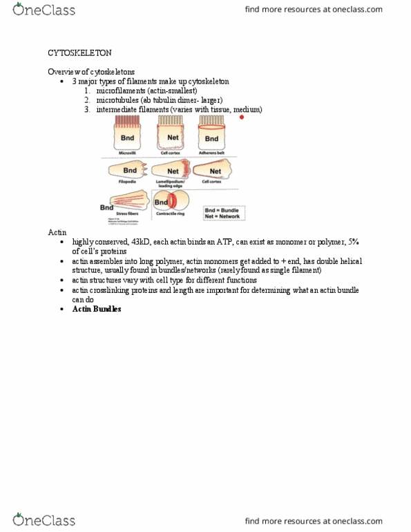
- Actin forms many important structures: these vary with cell type, so cells
can perform different functions
oActin bundles: found in microvilli, adherens belt, filopodia, stress
fibers, and contractile ring
Can be non-contractile: filaments crosslinked by fimbrin (short
crosslinking protein) to keep filaments straight and parallel. (+)
ends at one side and (-) ends at other side of cell. Filaments stay attached to
PM through lateral sidearm created by myosin I, allowing permanent
structure. Myosin II can’t fit in between filaments, so can’t contract
Ex: microvilli
oFound on epithelial cells of kidney or gut
oIncreases SA of PM for absorption
oFairly permanent structures
oAlso found on cells of inner ear (stereocilia that sense sound)
Stereocilia contain mechanically-gated channels that are bent by sound
waves. Bending pulls open channel on adjacent stereocilium, allowing K+
to flow in. Perceived as sound. Can break if sound wave too large
Can be contractile: filaments lined up anti-parallel to each other, joined
together using alpha-actinin (long crosslinking protein). Long crosslinker
allows myosin II to go in between filaments, allowing contractions to occur
Ex: Contractile ring
oUsed to divide cells in cytokinesis at the end of mitosis
oRing is a contractile actin bundle that
contains actin filaments and Myosin II
molecules
Ex: stress fibers
find more resources at oneclass.com
find more resources at oneclass.com

oused for keeping in touch with the surroundings. Just under the PM of a cell
that is sitting on a substrate (or dish)
oFocal adhesion: allows cell to adhere to
surrounding of cell. Found at both ends of a
stress fiber. Transmembrane proteins
(integrins) binds contractile bundles on one
side and ECM on other side
oActin networks: found in cell cortexes
Ex: cortical actin networks
supports the PM in all cells
In moving cells, it isn’t at the front of the leading edge
- Actin-based motility
oCells move using actin and Arp2/3 (actin related protein; always a dimer)
oMoving cells have 2 structures that use actin and Arp2/3 to help move
Filopodia: Arp2/3 complex nucleates actin filaments in filopodia,
allowing cell to move forward. Fimbrin keeps the filaments lined up
without myosin in between
Lamellipodium/leading edge: Arp 2/3 nucleates actin
polymerization in lamellipodia; causes branching at 70°
angles. Filamin crosslinks actin filaments to help keep
angle at 70°
oIntracellular bacterial infections
Actin and Arp2/3 hijacked by bacteria to allow invasion of neighboring
cells without bacteria being detected by immune system
Occurs in Listeria or Shigella bacteria
Bacteria taken in by phagocytosis, bacteria escape from phagosome, go
into cytoplasm, and Arp2/3 starts to nucleate actin
filaments at end, creating a plasma tail, pushing at cell’s
membrane. Can push hard enough to go into phagosome of
neighboring cell
Comet tail of actin pushes bacteria forward through the
cell: occurs through actin polymerization in the forward direction
- Myosin
oIn all cells
Myosin 1: attaches to actin filaments
Myosin 2: pulls actin filaments together
oIn some cells
Myosin VI and VII anchor stereocilia at their
base. If human is mutant for these, can cause deafness or blindness
- Summary
oTypes of crosslinkers
Crosslinkers for bundles
Fimbrin: in microvilli, filopodia, and focal adhesions
find more resources at oneclass.com
find more resources at oneclass.com

Alpha-actinin: in stress fibers and filopodia
Crosslinkers for networks
Filamin: at leading edges/lamellipodia
oStructures that contain actin
Average cells: not moving
Contain cortical network, stress fibers, and contractile rings when dividing
Moving cells
Contain cortical network, stress fibers (fewer), and contractile rings when
dividing
Also contain lamellipodia and filopodia
Cells with microvilli (epithelial, stationary)
Contain cortical network and have actin in their microvilli and other places
Hair cells in ear contain stereocilli and cortical network
oProteins involved with actin
Cross-linking proteins
Fimbrin, alpha-actin, filamin
Myosins
Myosin II: stress fibers, cortical networks (some), contractile ring
Myosin I: microvilli; attach membrane to side of actin filament
Myosin VI and VII: stereocilia contain them at sides and base of
actin bundles. Mutations cause deafness, if no stereocilia
Arp 2/3 complex
Lamellipodia: Arp2/3 nucleates and allows branching
Filopodia: Arp2/3 nucleates actin filaments in filopodia
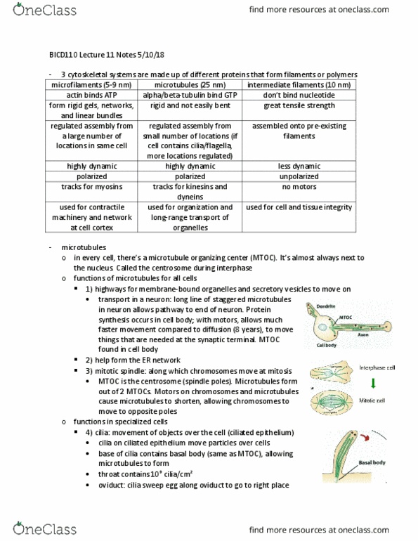
Lecture 11
microfilaments (5-9 nm) microtubules (25 nm) intermediate filaments (10 nm)
actin binds ATP alpha/beta-tubulin bind GTP don’t bind nucleotide
form rigid gels, networks,
and linear bundles
rigid and not easily bent great tensile strength
regulated assembly from
a large number of
locations in same cell
regulated assembly from
small number of locations (if
cell contains cilia/flagella,
more locations regulated)
assembled onto pre-existing
filaments
highly dynamic highly dynamic less dynamic
polarized polarized unpolarized
tracks for myosins tracks for kinesins and
dyneins
no motors
used for contractile
machinery and network
at cell cortex
used for organization and
long-range transport of
organelles
used for cell and tissue integrity
- 3 cytoskeletal systems are made up of different proteins that form filaments or polymers
find more resources at oneclass.com
find more resources at oneclass.com
Document Summary
The cytoskeleton: can be rigid or dynamic: keratocytes: skin cells on scales of fish. Cytoskeletons of the eukaryotic cell: 3 major types of filaments make up the cytoskeleton. Overview of actin: highly conserved, 43 kda, 5% of cell protein, can exist as a monomer or polymer, each actin binds an atp, in the cell, actin polymerizes into microfilaments/long polymers. There"s a (-) and (+) end, and actin monomers get added at the (+) end: has double helical structure. Rarely found as a single filament; instead, found in bundles or networks. Actin forms many important structures: these vary with cell type, so cells can perform different functions: actin bundles: found in microvilli, adherens belt, filopodia, stress fibers, and contractile ring. Can be non-contractile: filaments crosslinked by fimbrin (short crosslinking protein) to keep filaments straight and parallel. ends at one side and (-) ends at other side of cell. Pm through lateral sidearm created by myosin i, allowing permanent structure.