LIFESCI 4 Study Guide - Final Guide: Meiosis, Nonsense Mutation, Base Pair


1. Below are shown pedigrees from 3 families (labeled A, B and C), all of which has a
Klinefelter’s (XXY) male progeny. The DNA from each individual from each family was
digested with a restriction enzyme, run on a gel and then probed with a labeled X linked
piece of DNA that identifies two distinct RFLP morphs. The gel patterns for the parents
are shown below, however the banding pattern for the Klinefelter’s son is not shown. For
each pedigree draw the son’s banding pattern that would lead you to conclude that:
Family A had a non-disjunction event in meiosis 2 of the mother (there are no other
possibilities).
Family B had a non-disjunction event in meiosis 1 of the father or meiosis 1 of the
mother (there are the only two possibilities).
Family C had a non-disjunction event in meiosis 1 of the father (there are no other
possibilities).
find more resources at oneclass.com
find more resources at oneclass.com

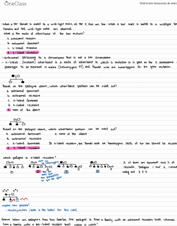
2. The human gene for hemophilia is on the X chromosome. Below is a pedigree from a
family afflicted with hemophilia. Blackened symbols indicate that the person has
hemophilia. To help with genetic diagnosis, a probe that detects an RFLP (restriction
fragment length polymorphism) on the X chromosome is used. This probe detects either a
7 kb restriction enzyme fragment or 3 kb and 4 kb restriction enzyme fragments. The
RFLP pattern for all the members of the pedigree is shown. The recombination distance
between the RFLP and the hemophilia locus is 15%. What information could be given to
the woman designated with the arrow as to the likelihood of her first son having
hemophilia? Show your work and circle your answer.
-15% chance she is a carrier;
-Therefore, 7.5% chance her son will have hemophilia
7 kb
4 kb
3 kb
find more resources at oneclass.com
find more resources at oneclass.com

3. Below is shown the RNA sequence from the imaginary protein coding region of the rII
gene of bacteriophage T4. As you can see the gene encodes a protein that is 7 amino
acids long. Met is the first amino acid. Previous analysis shows that the Val-Val-Val
amino acids at the C terminus of the protein (underlined below) are all that is required for
rII+ activity. You isolate 4 mutations in the gene. Mutation #1 and #2 are both positive
frameshift mutations, the base inserted is shown in bold. Mutations #3 and #4 are both
minus frameshift mutations, the base missing is shown as a gap in the sequence.
A. The following double mutants are made. For each double mutant write out the
sequence of the rII protein it will make. Predict (circle) whether the phage will be rII+ or
rII-.
B. You isolate an E. coli strain that contains a mutation in a tRNA gene. The normal
anticodon for this tRNA is 3' GCU 5', the mutated tRNA has the anticodon 3' ACU 5'.
This change in the anticodon does not affect the amino acid that attaches to the tRNA.
You infect this strain with wild type T4 and then look to see if an abnormal form of the
rII protein is made. If an abnormal form is made, what is its amino acid sequence.
Met ProGlyLysValValValArgTyr
find more resources at oneclass.com
find more resources at oneclass.com
Document Summary
Below are shown pedigrees from 3 families (labeled a, b and c), all of which has a. The dna from each individual from each family was digested with a restriction enzyme, run on a gel and then probed with a labeled x linked piece of dna that identifies two distinct rflp morphs. The gel patterns for the parents are shown below, however the banding pattern for the klinefelter"s son is not shown. For each pedigree draw the son"s banding pattern that would lead you to conclude that: Family a had a non-disjunction event in meiosis 2 of the mother (there are no other possibilities). Family b had a non-disjunction event in meiosis 1 of the father or meiosis 1 of the mother (there are the only two possibilities). Family c had a non-disjunction event in meiosis 1 of the father (there are no other possibilities): the human gene for hemophilia is on the x chromosome.