B STR 301 Study Guide - Final Guide: Vasoconstriction, Caput Medusae, Surface Tension

27 Jun 2018
School
Department
Course
Professor

WEEK 8 OBJECTIVES
1. Describe the gross and microscopic anatomy of the liver, gall
bladder, and bile duct system
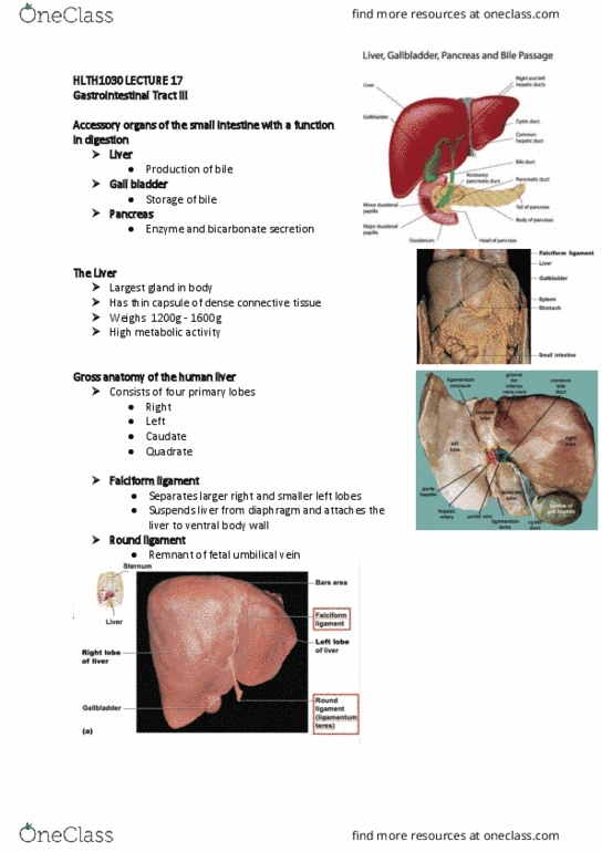
-Liver:
oGross anatomy:
Enclosed by a fibrous capsule
Serous membrane covers the capsule
Inferior to the diaphragm
Lobes (4):
Right (LARGEST)
Left
Quadrate
Caudate
Falciform ligament: separates right lobe from left lobe
Round ligament: remnant (leftover) of umbilical vein
find more resources at oneclass.com
find more resources at oneclass.com

WEEK 8 OBJECTIVES
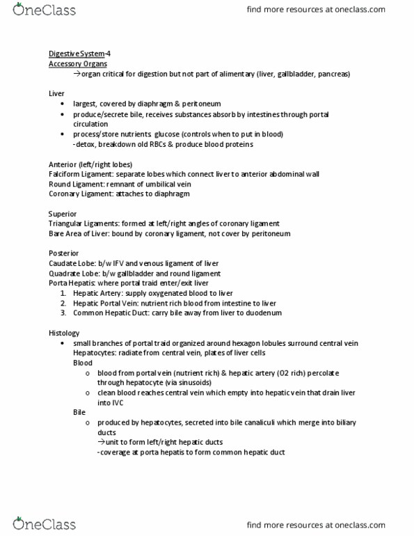
oMicroscopic Anatomy:
Liver tissue made up of units called hepatic lobules
Hepatic lobules: small cylinders with central vein, radiating plates of
hepatocytes
Hepatocyte: liver cells
oProduces and secretes bile into the bile canaliculi
Bile drains into the bile ducts
Bile flows in the opposite direction of blood
No mixing of bile and processed blood
Plates of hepatocytes = yellow
Hepatic sinusoids: leaky capillaries between plates of hepatocytes
Located between the radiating plates
Triads: at the corners of the lobules
Hepatic portal vein branch
Hepatic artery branch
Bile duct
The blood from the hepatic portal vein branch will combine with the
blood from the hepatic artery branch
Oxygenated blood mixing with deoxygenated blood
This blood will dump into the central vein of the lobule
oBlood is already processed by the hepatocyte at this
point when it gets to the central vein
Remove toxins
add blood proteins
other liver functions
owill then drain from the liver dump into inferior vena
cava heart
Hepatic (liver) macrophages: remove bacteria and other microbes in
the blood that enter the liver
Pink things in the image below
oHepatic Portal System: brings blood from the digestive tract (rich in
nutrients) drains into the hepatic portal vein liver
Delivers nutrient rich blood to liver for processing!!!
SUMMARY:
Hepatic portal vein: brings nutrient-rich blood from veins of
GI tract to liver
Hepatic artery: brings arterial blood
oAorta celiac trunk common hepatic artery hepatic
artery proper hepatic artery
Hepatic veins: exit from the top of liver and empty into
inferior vena cava
find more resources at oneclass.com
find more resources at oneclass.com

WEEK 8 OBJECTIVES
-GALLBLADDER:
oSac on the underside of liver
oStores and concentrates bile
oCystic duct
Carries bile to bile duct
-BILE PASSAGEWAYS:
oTwo hepatic ducts merge to form the common hepatic duct
oCommon hepatic duct merges with cystic duct to form bile duct
oBile duct merges with main pancreatic duct hepatopancreatic ampulla at
major duodenal papilla
Combining of bile from bile duct and pancreatic duct
find more resources at oneclass.com
find more resources at oneclass.com
Document Summary
Week 8 objectives: describe the gross and microscopic anatomy of the liver, gall bladder, and bile duct system. Falciform ligament: separates right lobe from left lobe. Round ligament: remnant (leftover) of umbilical vein. Liver tissue made up of units called hepatic lobules. Hepatic lobules: small cylinders with central vein, radiating plates of hepatocytes. Hepatocyte: liver cells: produces and secretes bile into the bile canaliculi. Bile flows in the opposite direction of blood. No mixing of bile and processed blood. Hepatic sinusoids: leaky capillaries between plates of hepatocytes. The blood from the hepatic portal vein branch will combine with the blood from the hepatic artery branch. This blood will dump into the central vein of the lobule: blood is already processed by the hepatocyte at this point when it gets to the central vein. Other liver functions: will then drain from the liver dump into inferior vena cava heart.