HPHY 322 Study Guide - Final Guide: Resting Potential, Fovea Centralis, Transducin

4 Dec 2015
School
Department
Course
Professor
Document Summary
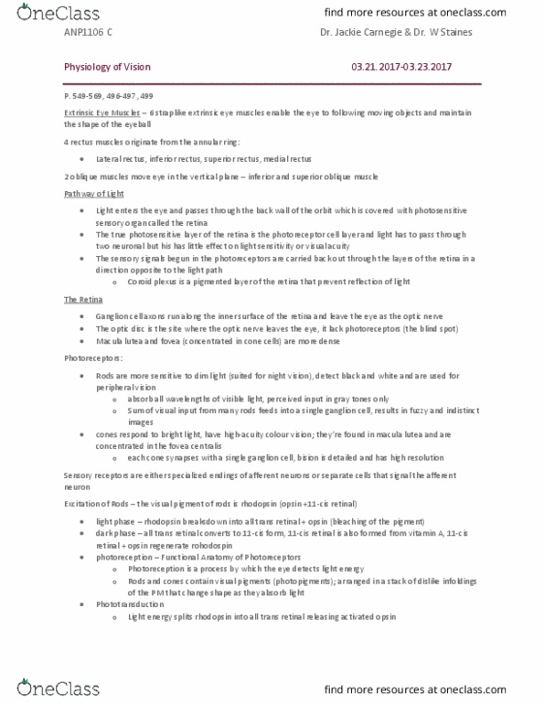
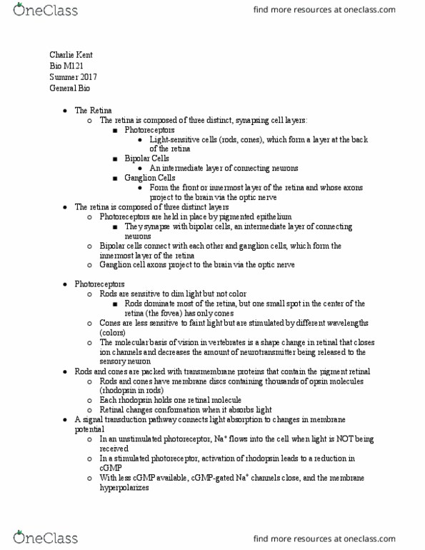
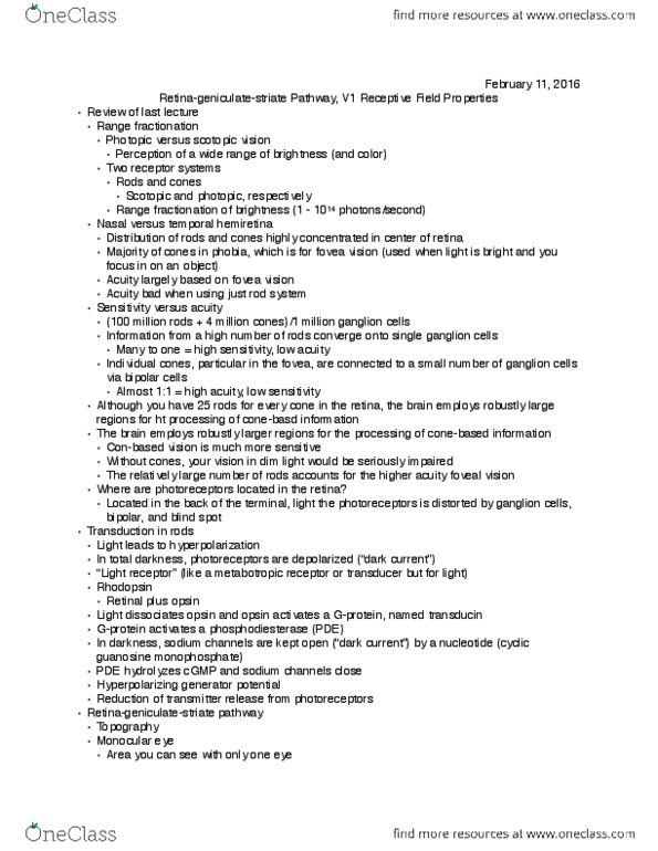
Rods - dim light, black and white, located in the periphery of the retina. Cones - color, color pigment, located in the fovea centralis (densely packed) Melanin: prevents reflection and absorbs light animals have low melanin humans have high melanin. Pde breaks down (hydrolyzes) camp and cgmp. What happens to rhodopsin when it absorbs light? retinal detaches from rhodopsin and rhodopsin becomes activated which activates the g protein transducin. How does activated rhodopsin affect cgmp? activated rhodopsin activates g protein transducin which activates pde which breaks down the concentration of cgmp in the rod cell. How does cgmp affect sodium ion channels? decrease in cgmp causes the closure of cgmp gated na+ and ca2+ ion channels so cation influx does not occur. How does hyperpolarization affect glutamate release? hyperpolarization decreases the release of glutamate neurotransmitter. Which cells have action potentials? only ganglion cells which their axons make up the optic nerve.