PSYC 372 Study Guide - Final Guide: Magnetic Resonance Imaging, Positron Emission Tomography, Myelin

4 May 2018
School
Department
Course
Professor

Final Exam Review
Cumulative Material
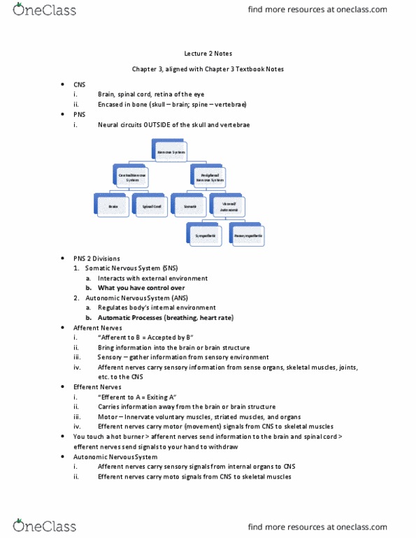
1. What are the two major divisions of the nervous system? Where in the body is each division
located?
a. The Central Nervous System (CNS): Brain, Spinal Cord, Retina of the Eye
i. Encased in bone (skull – brain; spine – vertebrae)
2. The Meninges: 3 protective membranes enclosing the brain and spinal cord
a. Dua Mate: had othe losest to skull/spie
b. Arachnoid layer: Spider web-like middle layer
❖ Contains the subarachnoid space filled with any blood vessels
❖ Contains the cerebrospinal fluid (CSF)
c. Pia Mate: pious/soft othe that adhees to sufae of ai ad spial od
3. What is the function of the ventricular system? What are the ventricles of the brain?
a. The ventricular system produces cerebrospinal fluid (CSF) and cushions the CNS
❖ The fluid flow from the lateral ventricle > 3rd ventricle > cerebral aqueduct
> 4th ventricle > travels down the spinal cord and brain until it is reabsorbed
b. The Ventricles of the Brain
i. Lateral ventricles (ventricles 1 and 2) (two halves of the brain)
ii. 3rd ventricle (in-between)
iii. 4th ventricle (smallest one)
4. Describe the functions of all these neuron parts AND be able to label them on a graph
a. Cell od: ai of the euo; etaoli ete of the euo; otais the ogaelles
(nucleus, mitochondria, ribosomes), also contains DNA
b. Cell eae: oudaies, seipeeale eae that eloses the euo
i. Composed of a lipid bilayer (2 layers of fat molecules)
ii. Hydrophobic region (non-pola tails ate feaig
iii. Hydrophilic region (polar head uses hdoge ods to get lose to ate ate
loig
iv. Proteins are stuck within the lipid bilayer; however, they are not stationary, they are
moving around – going in and out of the cell
c. Dendrites: Short branches extending from the cell body; receptors on the surface receive
information from other neurons, bringing information into the cell
d. Axon Hillock: junction between the cell body and the axon; starting location of the action
potential
e. Ao: ifoatio ete ad ifoatio sede of the euron; transmits electrical
impulses
i. Myelin sheath: fatty whitish-gray substance insulating the axons; speeds up
conduction of a signal down the axon
ii. Nodes of Ranvier: allows the signal to jump from node to node (saltatory
conduction)
f. Terminal Buttons: endpoints of axon branches, site of release for chemical signals that will
reach other neurons
find more resources at oneclass.com
find more resources at oneclass.com

5. What is the function of the myelin sheath? What are the nodes of Ranvier, and how do they
contribute to saltatory conduction of an electrical signal down an axon?
a. The myelin sheath is the fatty whitish-gray substance that insulates the axons and speeds
the conduction of a signal down the axon
b. Nodes of Ranvier allow the signal to jump from node to node
i. Increases the conduction velocity of action potentials
6. Describe the functions of the following glial cells:
a. Microglial: small scavenger cells that respond to injury or disease in the CNS; contain
phagocytes that destroy viruses/bacteria/microorganisms; go from dormant to activated
state in the presence of CNS infection of inflammation
b. Oligodendrocytes/Schwann Cells: build the myelin sheaths that wrap around axons of
certain CNS neurons; one oligodendrocyte for many axons; Schwann cells have the same
function but are only found in the PNS; one Schwann cell for one axon
c. Astrocytes: Star-shaped cells; cover outer surfaces of blood vessels in the brain and assist
with blood-brain function
d. Radial Glia: guide migration and growth of neurons during brain development; later become
neurons or a different type of glial cell
7. Name the 5 subdivisions of the brain that form the 3 major divisions (forebrain, midbrain, and
hindbrain)
a. Forebrain: telencephalon (thalamus, hypothalamus), diencephalon (cerebral cortex and
limbic system)
b. Midbrain: mesencephalon (tectum)
c. Hindbrain: myelencephalon (the medulla) and metencephalon (pons and cerebellum)
8. Name the four lobes of the cerebral cortex and their main functions/important brain regions
within them.
a. Frontal lobe, parietal lobe, occipital lobe, and temporal lobe
9. What is membrane potential?
a. Difference in electrical charge between the inside and the outside of a cell
b. Scientists first used the axon of the squid because the axon extends from the head to the
tail of the squid which made it easier to study
10. What is resting potential? What does it mean to say that a neuron is polarized when it is at rest?
a. The value of the membrane potential in a neuron that is at rest
b. When a neuron is polarized at rest that means the inside of the neuron is negatively charged
relative to the extracellular fluid
11. Name the 4 types of ions (excluding calcium) who distribution inside vs. outside of a neuron
contributes to the resting potential. Which are anions, and which are cations? Which are found
at great concentrations inside the cell, and which are found in greater concentrations outside
the cell?
a. Sodium (Na+ and OUTSIDE THE CELL)
b. Potassium (K+ and INSIDE THE CELL)
c. Chloride (Cl- and OUTSIDE THE CELL)
d. Calcium (Ca++ and outside the cell)
e. Ogai aios egatiel haged ad stuk iside the ell eause the ae e large
ad soeties at e taspoted
find more resources at oneclass.com
find more resources at oneclass.com

12. How does a sodium-potassium pump work? Which ions does it pump in and out of the cell and
what amount?
a. An active transport mechanism; contributes to the uneven distribution of ions at rest
b. Transports THREE SODIUM IONS (Na+) OUT OF THE CELL for every TWO POTASSIUM (K+)
IONS it brings INTO THE CELL
13. Be able to draw and explain the steps of the action potential
a.
14. What is the absolute and relative refractory periods?
a. Absolute refractory period: 1-2 milliseconds after an action potential has been fired, it is
impossible to trigger another one
b. Relative refractory period: For a few more milliseconds, an action potential can only be fired
by applying higher than normal amounts of stimulation
15. What is the difference between an EPSP and an IPSP? How does each of these affect the
likelihood that a postsynaptic neuron will fire an action potential?
a. Depolarizations are excitatory postsynaptic potentials (EPSPs): they INCREASE the likelihood
that the postsynaptic neuron will fire an action potential
b. Hyperpolarizations are inhibitory postsynaptic potentials (IPSPs): the DECREASE the
likelihood that the postsynaptic neuron will fire an action potential
16. Types of structural imaging, functional imaging, and noninvasive stimulation
a. Computerized Tomography (CT) Scanning
i. Structural Imaging; a contrast x-a tehiue ai tissue doest sho up
ii. Inject a substance (iodine) that absorbs x-rays to enhance contrast and allow
visualization of brain tissue
b. Magnetic Resonance Imaging (MRI)
i. Structural Imaging; determines the amount of certain types of atoms in different
locations in the body; generates a series of 2D or 3D images
ii. Static magnetic field > radiofrequency pulse
c. Advantages of MRI over CT
i. No exposure to radioactivity is required
ii. Images generated have better resolution
d. Positron Emission Tomography (PET)
i. Functional Imaging; 2D maps of radioactivity rather than detailed images of the
brain
ii. Inject radioactive 2-DG ito the patiets aotid ate
e. Functional MRI (fMRI)
find more resources at oneclass.com
find more resources at oneclass.com
Document Summary
Where in the body is each division located: the central nervous system (cns): brain, spinal cord, retina of the eye. Encased in bone (skull brain; spine vertebrae: the meninges: 3 protective membranes enclosing the brain and spinal cord, du(cid:396)a mate(cid:396): (cid:862)ha(cid:396)d (cid:373)othe(cid:396)(cid:863) (cid:272)losest to skull/spi(cid:374)e, arachnoid layer: spider web-like middle layer. Contains the subarachnoid space filled with any blood vessels. What are the ventricles of the brain: the ventricular system produces cerebrospinal fluid (csf) and cushions the cns. The fluid flow from the lateral ventricle > 3rd ventricle > cerebral aqueduct. > 4th ventricle > travels down the spinal cord and brain until it is reabsorbed: the ventricles of the brain. Lateral ventricles (ventricles 1 and 2) (two halves of the brain) Composed of a lipid bilayer (2 layers of fat molecules) Hydrophilic region (polar head uses h(cid:455)d(cid:396)oge(cid:374) (cid:271)o(cid:374)ds to get (cid:272)lose to (cid:449)ate(cid:396)(cid:895) (cid:862)(cid:449)ate(cid:396) lo(cid:448)i(cid:374)g(cid:863)