BPK 105 Study Guide - Midterm Guide: Uvea, Retina, Ciliary Muscle

27 Apr 2018
School
Department
Course
Professor

Module 5 - Objectives - Part 3
● Describe how light is focused on the retina including the functions and
relevant structures of each tunic (layer) in the eye.
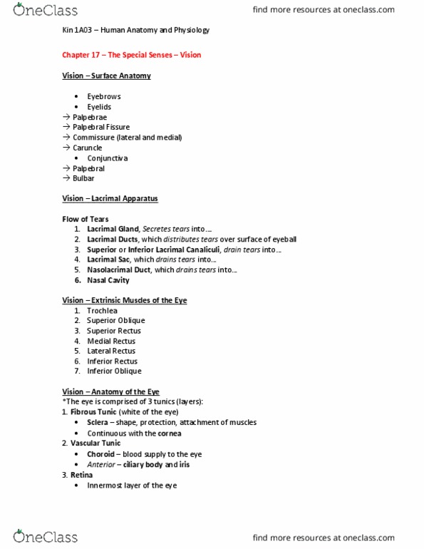
Eyeball: a hollow, fluid-filled sphere.
- wall of the eye is composed of three tissue layers/tunics (figure 9.9).
- outer, fibrous tunic consists of the sclera and cornea.
- middle, vascular tunic consists of the choroid, ciliary body, and iris.
- inner nervous tunic consists of the retina.
Fibrous Tunic
-Sclera: firm, white, outer connective tissue layer of the posterior five-sixths of the fibrous
tunic.
- helps maintain the shape of the eye, protects the internal structures, and provides
attachment sites for the extrinsic eye muscles.
- small portion of the sclera can be seen as the “white of the eye.”
-Cornea: the transparent anterior sixth of the eye, which permits light to enter.
- As part of the focusing system of the fibrous tunic, the cornea also bends, or refracts, the
entering light.
Vascular Tunic
- middle tunic of the eye
- contains most of the blood vessels of the eye.
- posterior portion of vascular tunic, associated with the sclera, is the choroid ←- thin
structure consists of a vascular network and many melanin-containing pigment cells,
causing it to appear black. The black color absorbs light, so that it is not reflected inside
the eye. (If light were reflected inside the eye, the reflection would interfere with vision.)
- Anteriorly, vascular tunic consists of the ciliary body and the iris.
-ciliary body = continuous with the anterior margin of the choroid.
- contains smooth muscles called ciliary muscles, which attach to the perimeter of the
lens by suspensory ligaments (figure 9.10).
-lens is a flexible, biconvex, transparent disc (see figure 9.9).
-iris = the colored part of the eye.
Document Summary
Describe how light is focused on the retina including the functions and relevant structures of each tunic (layer) in the eye. Eyeball : a hollow, fluid-filled sphere. wall of the eye is composed of three tissue layers/ tunics (figure 9. 9). outer, fibrous tunic consists of the sclera and cornea. Middle, vascular tunic consists of the choroid, ciliary body, and iris. inner nervous tunic consists of the retina. Cornea : the transparent anterior sixth of the eye, which permits light to enter. As part of the focusing system of the fibrous tunic, the cornea also bends, or refracts, the entering light. The black color absorbs light, so that it is not reflected inside the eye. (if light were reflected inside the eye, the reflection would interfere with vision. ) Light passes through the pupil, and the iris regulates the diameter of the pupil , which controls the amount of light entering the eye.