BIOL 330 Study Guide - Final Guide: Endonuclease, Telomerase, Base Pair

23 Feb 2017
School
Department
Course
Professor
Document Summary
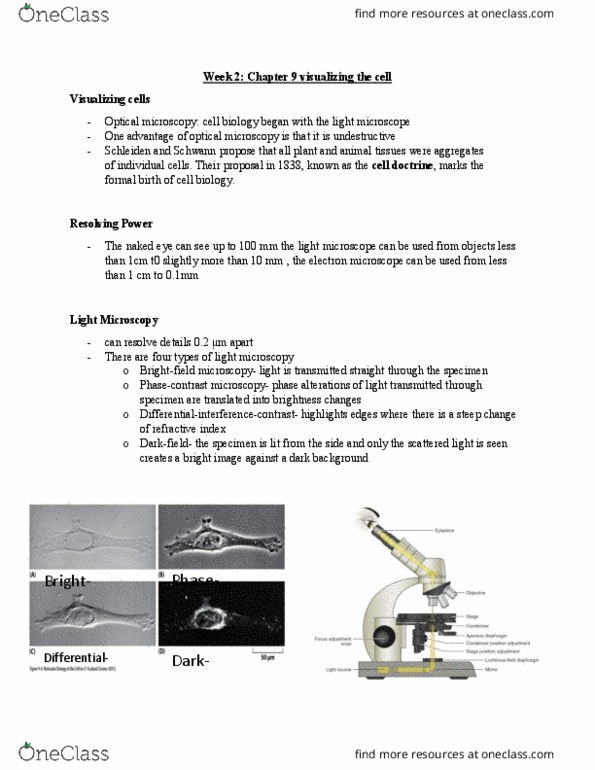
Resolving power: bright-field- in which light is transmitted straight through the specimen. Bottom- light is coming from a ring and shining through the tissue: phase-contrast microscopy- in which phase alterations of light transmitted through the specimen are translated into brightness changes. Used to look at alive cells (not fixed or frozen) Differential-interference-contrast microscopy: highlights edges where these is a steep change of refractive index. Inject target protein into animal which then makes specific antibodies to your protein: can only see the commercially-made secondary antibody which bind primary antibodies, secondary antibody recognizes the common part of the primary antibody. Step 1- an unlabelled primary antibody is added and targets a certain antigen. Step 2- a secondary labelled antibody is added that targets the primary antibody. This microscope is similar to an ordinary light microscope except that the illuminating light is passed through two sets of filters. Most often used to detect specific proteins or other molecules in cells and tissues.