MEDRADSC 2H03 Study Guide - Midterm Guide: Superior Mesenteric Vein, Peptic Ulcer, Duodenal Bulb

2 Feb 2017
School
Department
Course
Professor
Document Summary
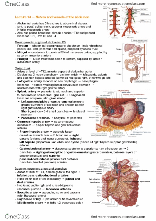
The stomach is located under the left dome of the diaphragm, with the superior part joining the esophagus at the cardiac sphincter, creating the gastroesophageal junction. Rounded top is the fundus, which touches the diaphragm superiorly. Anterior surface in contact with the anterior abdominal wall, left lobe of the liver and the diaphragm. Posterior is the gastric portion of the spleen, the left adrenal gland and kidney and the body/tail of the pancreas. Arterial blood is supplied by branches of the gastric, splenic and gastroduodenal arteries. The gastric veins usually drain directly into the portal vein or into the superior mesenteric vein. Pancreas, the duodenum has a loop and the head the pancreas sits in the c-part. 1st portion: superior, has duodenal bulb, most common site for duodenal ulcers. 2nd portion: descending, most lateral part, sphincter of oddi, ampulla of vater. The ileum is the lower 3/5ths of the small bowel.