BIOLOGY 2B03 Study Guide - Final Guide: Electron Microscope, Eyepiece, Vacuole


Document Summary
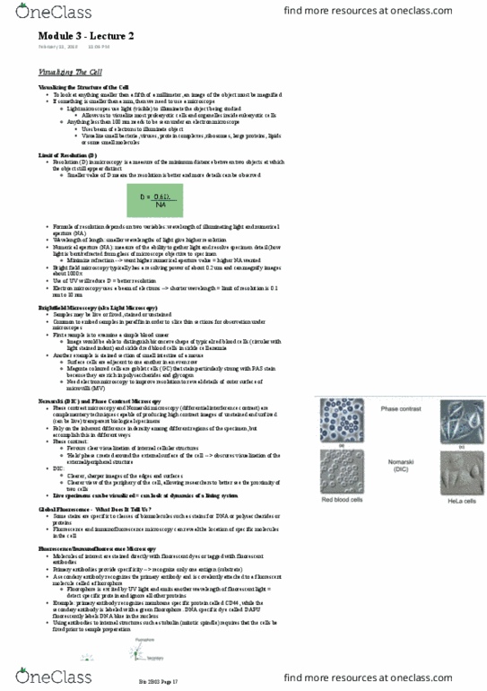
Red = phase contrast and differential interference contrast (dic) microscopy video ( "s notes) Blue = preparation of embryos for electron microscopy of video ( "s notes) Resolving objects that are less than 200 nm in size (0. 2 um)- live cells and tissues that lack compounds that absorb light (need to stain them) Contrast is generated by differences in index of refracting of object, and its surrounding medium. Images appear to cast a shadow to one side = represents difference in refractive index of specimen rather than its topography. In addition to brightfield: polarizer between light source and condenser. Takes advantage of differences in refractive index and thickness of cellular materials. Produce images that differ in appearance and reveal diff. features of cell architecture. Entire object + subcellular structures are highlighted by interference rings = concentric halos of dark and light bands. Tracking location of a small bead of known size to a precision of a few nm.