BCH3052 Final: BCH3052 Exam Notes

25 May 2018
School
Department
Course
Professor
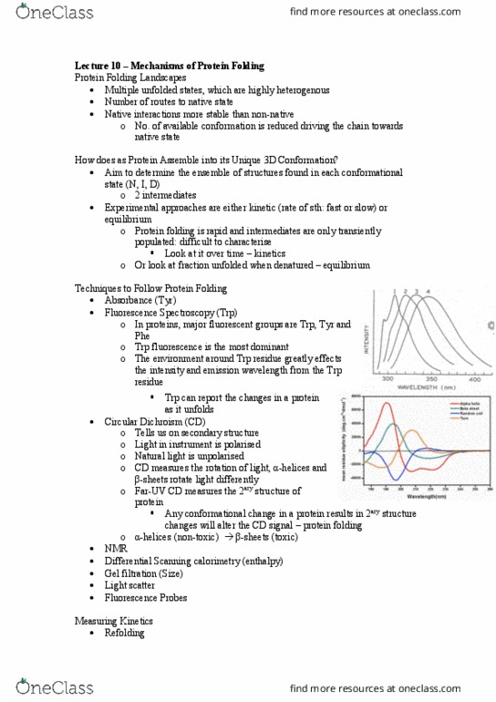
BCH-3052 NOTES
Semester 2
Exam Styles (2 hours)
• 60%
• Short Answer Questions (7 questions – 15 minutes each)
o Answer 1 short question from a choice of 2 questions for each section
o Section A: Jackie Wilce (Protein Structure)
! Lectures 1-4
o Section B: Matt Wilce
! Lectures 5-7
o Section C: Mibel Aguilar (Protein Folding & Misfolding)
! Lectures 8-12
o Section D: Mibel Aguilar (Proteomics)
! Lectures 13-15
o Section E: Jackie Wilce (NMR and Protein Engineering)
! Lectures 16-18
o Section F: Anna Roujeinikova
! Lectures 23-24
o Section G: Martin Stone
! Lectures 19-22
UNIT Objectives
• Describe the relationship between protein sequence, structure and function and
relate this to specific examples in human health and disease;
• Explain how proteins fold to their correct three dimensional shape and how
this process may go wrong and cause disease;
• Illustrate how NMR and X-ray crystallography are used to determine the
structure of a peptide or protein;
• Critically analyse how our understanding of proteins contributes to
biotechnology and medicine, in particular comment on the contribution of
protein engineering and proteomics to these fields;
• Describe the use of fluorescent proteins as tools for characterising the role of
proteins in vivo;
• Apply experimental techniques and methodologies to determine the structure
and function of an unknown protein.

BCH3052 – Advanced Protein Biology
Jackie Wilce
Introduction and Conformational Change in
Proteins
Lecture 1 – Introduction to Tertiary Structures
of Proteins
• Primary structure: amino acids linked by
covalent bonds
• Secondary Structure
o Alpha helix: 3.6 aa per turn
! Bond angles of rotation are optimal and bonding patterns
stabilise the structure
! Hydrogen bonds holding stabilise alpha helix
! Proline doesn’t have NH – alpha helix breaker
o Beta turn
! Carbonyl of aa 1 forms hydrogen bond with aa 4
o Beta sheets
! Antiparallel
• Tertiary Structure
o Determination of 3D structures of proteins using X-ray crystallography
! Protein needs to be crystalized " X rays bounce of repeating
electron patterns " interpreted to recreate maps of electron
density
o OR 2D NMR
Calcium Signalling
• External signals from receptors " results in release of calcium into cytoplasm
of cells
• Intracellular calcium fluxes are rapid " result in oscillations
• Many proteins turn on after calcium flux
• E.g. Calmodulin – protein conformational change controlled by calcium ions
! Short chain of amino acids
! EF hands - Helix turn helix motif
! Helix turn helix EF hand
! Helix turn helix
o Each loop contains calcium binding site
o Binding site of calmodulin are hydrophobic alpha helixes of target
proteins
Document Summary
Exam styles (2 hours: 60, short answer questions (7 questions 15 minutes each, answer 1 short question from a choice of 2 questions for each section, section a: jackie wilce (protein structure) Lectures 1-4: section b: matt wilce. Lectures 5-7: section c: mibel aguilar (protein folding & misfolding) Lectures 8-12: section d: mibel aguilar (proteomics) Lectures 13-15: section e: jackie wilce (nmr and protein engineering) Lectures 16-18: section f: anna roujeinikova. Lectures 23-24: section g: martin stone. Lecture 1 introduction to tertiary structures of proteins: primary structure: amino acids linked by covalent bonds, secondary structure, alpha helix: 3. 6 aa per turn. Bond angles of rotation are optimal and bonding patterns stabilise the structure. Proline doesn"t have nh alpha helix breaker: beta turn. Carbonyl of aa 1 forms hydrogen bond with aa 4: beta sheets. Antiparallel: tertiary structure, determination of 3d structures of proteins using x-ray crystallography.