HEALTH EDUCATION Lecture Notes - Rectus Capitis Posterior Minor Muscle, Rectus Capitis Posterior Major Muscle, Suboccipital Nerve

20 Oct 2022
Department
Course
Professor
Document Summary
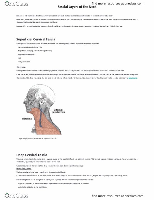
The suboccipital muscles are a group of four muscles situated underneath the occipital bone. All the muscles in this group are innervated by the suboccipital nerve. They are located within the suboccipital compartment of the neck; deep to the sternocleidomastoid, trapezius, splenius and semispinalis muscles. They collectively act to extend and rotate the head. In this article, we shall look at the anatomy of the suboccipital muscles their attachments, actions and innervation. The rectus capitis posterior major is the larger of the rectus capitis muscles. It is located laterally to the rectus capitis posterior minor. Attachments: originates from the spinous process of the c2 vertebrae (axis), and inserts into the lateral part of the inferior nuchal line of the occipital bone. The rectus capitis posterior minor is the most medial of the suboccipital muscles. There is a connective tissue bridge between this muscle and the dura mater (outer membrane of the meninges) which may play a role in cervicogenic headaches.