NURS 472 Lecture Notes - Lecture 2: Atrioventricular Node, Hypokalemia, Evry

16 Jan 2018
School
Department
Course
Professor
Document Summary
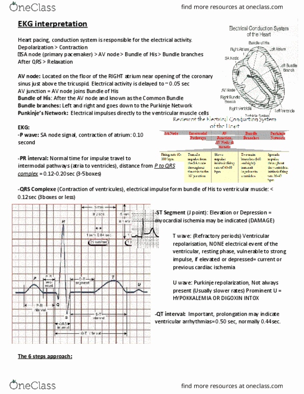
Aortic uses receptors in the carotid sympathetic kicks in. Bainbridge uses receptors in the vena cava parasympathetic kicks in. Monitoring lead systems 3 electrode and 5 electrode (5 is what we use on cvsu) If dysrhythmia you run a 12 lead ekg. Straight line on monitor 1st thing you do is check the patient (usually leads are off) Horizontal small square is equal to 0. 04 seconds. Each large square (5 small squares) is 0. 2 seconds. Vertical marking distances are 3 seconds usuall(cid:455) do(cid:374)"t ha(cid:448)e a u--- u usually indicates electrolyte imbalance but can occur in healthy patients: p wave. Depolarization of the atria; normally all should look alike and one p wave should precede every qrs: pr interval --- beginning of atrial contraction to beginning of ventricular contraction. Onset of atrial depolarization to the onset of ventricular depolarization; measure from beginning of p wave to beginning of qrs complex. Should be regular and 0. 12-0. 20 seconds: qrs complex.