BIOL 206 Lecture Notes - Lecture 6: Cell Wall, Sieve Tube Element, Casparian Strip

Document Summary
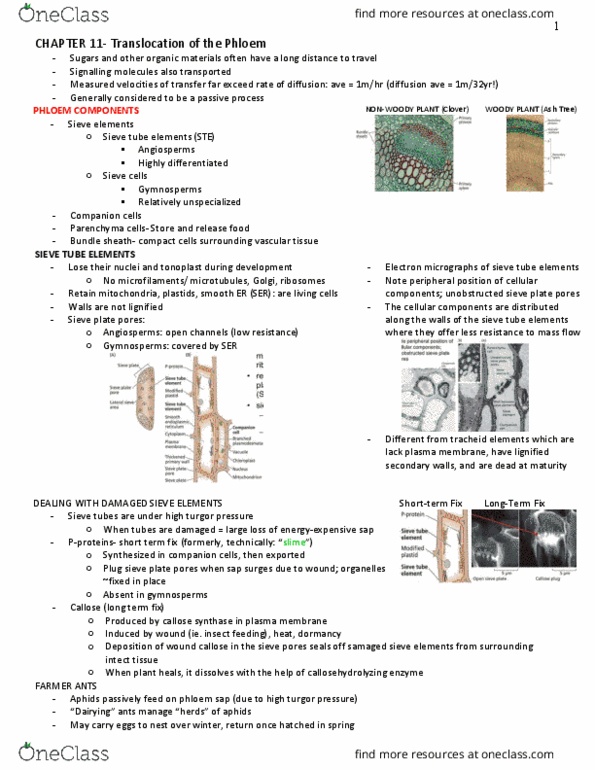
Parenchyma alive, thin primary cell walls, contain living protoplast and large vacuole for storage. Can be various shapes and sizes depending on what tissue it is located in. Each tube element is closely associated with one or more specialized precnchyma cell, known as companion cells. Companion cells contain a nucleus (unlike sieves) and supply many vital macromolecules and metabolites to the sieve tube via the abundant plasmodesmata that connect them. Collenchyma alive, possess a primary cell wall that is non-uniform and typically thickened in the corners. Several different types depending on the pattern of cell wall thickening. Main function is to provide support and protection in tissues that are growing rapidly (by way of their thickened primary cell walls) such as leaf stalks of celery and rhubarb. Sclerenchyma dead, very thick secondary cell walls that are typically lignified (cell wall polysaccharides and proteins interlinked by a tough, degradation-resistant phenolic polymer known as lignin).