PSYCH 454 Lecture Notes - Lecture 9: Electromagnetic Spectrum, Extraocular Muscles, Electromagnetic Radiation

24 May 2018
School
Department
Course
Professor

Lecture 9: General Concepts Eye/Light
Light
● Electromagnetic wave
●Wavelength- one cycle of wave; trough to trough or peak to peak
● Electromagnetic spectrum- different wavelengths of light
○ High energy gamma rays
○ Lower energy
○ Visible spectrum (we can see) forms a narrow band between UV and Infrared
ray→ visible range: 400 nm - 700 nm
■ 400 nm → high energy, blue
■ 700→ lower energy, red
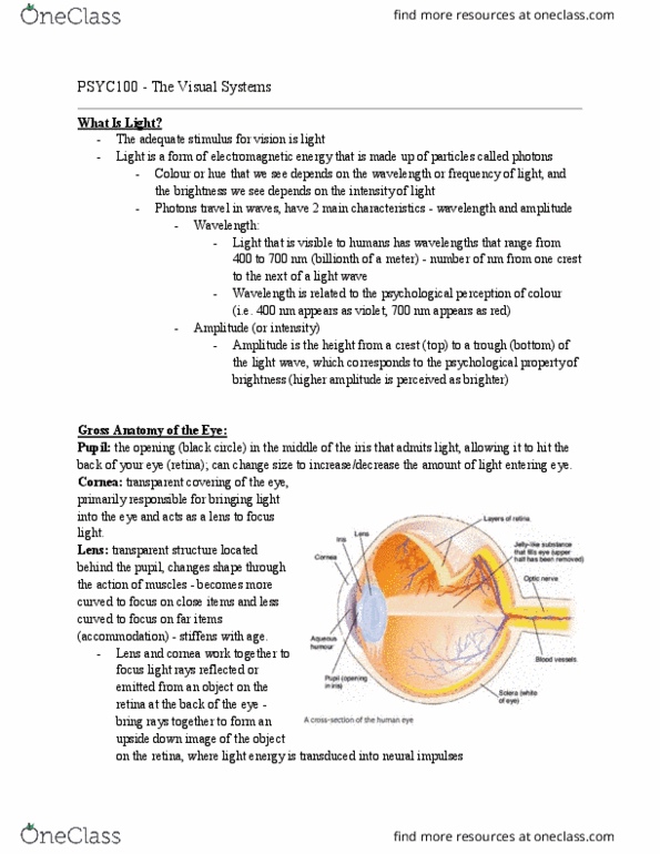
Gross Anatomy of the Eye
● Pupil: aperture of eye; light comes through pupil into eye
● Iris: controls diameter of pupil; controls amt of light entering eye
● Extraocular muscles: 6, allow to move eye
● Optic nerve: back of eye, info from retina to brain
●
● Light comes through cornea on surface of eye, focused by lens on back of the eye
○ Back of eye: retina→ has photosensitive cells and processing visual scene cells
■ Most sensitive part of retina: fovea → if your focusing on an object, this is
where light hits!
● Blood vessels
● Optic nerve and blood vessels pass through optic disk
Structure of the Retina
●Fovea: most sensitive part of retina; in central part of retina
○ Receives light from the center of the visual field; receives light where focused
○ Anything to right of fovea (dashed line): temporal retina
○ Left of dashed line: nasal retina
Document Summary
Wavelength - one cycle of wave; trough to trough or peak to peak. Visible spectrum (we can see) forms a narrow band between uv and infrared ray visible range: 400 nm - 700 nm. Pupil: aperture of eye; light comes through pupil into eye. Iris: controls diameter of pupil; controls amt of light entering eye. Extraocular muscles: 6, allow to move eye. Optic nerve: back of eye, info from retina to brain. Light comes through cornea on surface of eye, focused by lens on back of the eye. Back of eye: retina has photosensitive cells and processing visual scene cells. Most sensitive part of retina: fovea if your focusing on an object, this is where light hits! Optic nerve and blood vessels pass through optic disk. Fovea: most sensitive part of retina; in central part of retina. Receives light from the center of the visual field; receives light where focused. Anything to right of fovea (dashed line): temporal retina.