PSYCH 454 Lecture Notes - Lecture 19: Supplementary Motor Area, Somatotopic Arrangement, Posterior Parietal Cortex

24 May 2018
School
Department
Course
Professor

Lect 19: Central Motor System
Cortical areas contributing to control of Voluntary
Movements
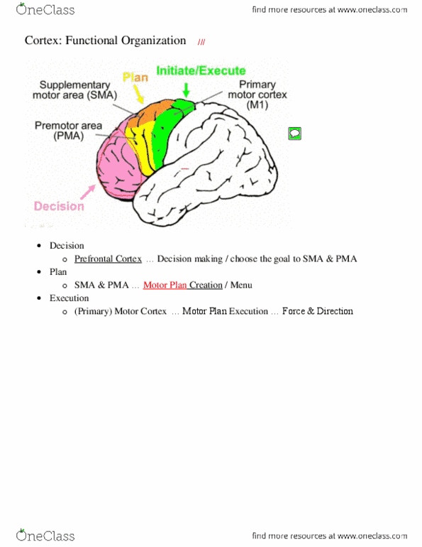
●Motor areas→ areas 4 and 6 (anterior of central
sulcus)
○M1- primary motor area
○PMA- premotor area
○SMA- supplementary motor area
●Prefrontal and posterior parietal cortex (area 5/7)
connect with area 6
○Ex: cognitive control signals sent to area 6
(SMA, PMA)
●Area 6: contributes to motor planning
●Areas 4 and 6 contribute to most axons to corticospinal tract
○For motor commands to be sent to motor units
●Somatosensory (area 5,7) → interact with motor cortex and associated areas to help
guide action
Somatotopic map in Primary Motor Cortex (M1) (classic view)
●Brief M1 electric stimulation → causes twitch of particular contralateral
muscles
■Ex: stimulate Left motor cortex in facial region on map → get a twitch of
right facial muscle
●As you stimulate different M1 sites, different muscles = affected
●Homunculus man map→ greater regions for hands, face, tongue (less for trunk, lower
extremities)
Alternative Hypothesis: Motor Cortex organized into action zones?
●Challenges classical view of somatotopic map of M1 (above)
●Longer M1 electrical stimulation causes more complex movements
○Stimulating for 500 ms (rather than 50 ms) more similar to usual duration of M1
firing
■Not just a twitch→ cause MORE COMPLEX movements + full actions (ex:
clapping, or feeding movements)
■Mimics natural firing of classic map
●Action zone: parts in motor cortex, represents a pattern of motor cortex that you can
stimulate for long period of time that will elicit complex movements
●Organization of action zones = consistent with somatotopic map (classical view)
○Dorsal portion of motor cortex → manipulations in hands
Document Summary
Motor areas areas 4 and 6 (anterior of central sulcus) Prefrontal and posterior parietal cortex (area 5/7) connect with area 6. Ex: cognitive control signals sent to area 6 (sma, pma) Areas 4 and 6 contribute to most axons to corticospinal tract. Somatosensory (area 5,7) interact with motor cortex and associated areas to help. For motor commands to be sent to motor units guide action. Somatotopic map in primary motor cortex (m1) (classic view) Brief m1 electric stimulation causes twitch of particular contralateral muscles. Ex: stimulate left motor cortex in facial region on map get a twitch of right facial muscle. As you stimulate different m1 sites, different muscles = affected. Homunculus man map greater regions for hands, face, tongue (less for trunk, lower extremities) Challenges classical view of somatotopic map of m1 (above) Longer m1 electrical stimulation causes more complex movements. Stimulating for 500 ms (rather than 50 ms) more similar to usual duration of m1 firing.