BIOL 212 Lecture Notes - Lecture 20: Photographic Film, Radiodensity, Calcification

5 Aug 2020
School
Department
Course
Professor
Document Summary
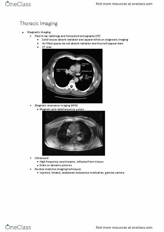
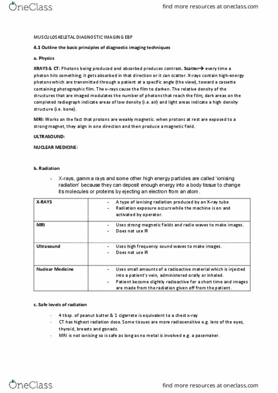
Procedures and techniques of radiology and diagnostic imaging. Noninvasive ultrasound of the kidneys that is useful in distinguishing between fluid-filled cysts and solid masses, detecting renal calculi, identifying obstructions, and evaluating transplanted kidneys. Ultrasound examination that"s important in distinguishing solid thyroid nodules from cystic nodules. Technique used to prepare an x-ray image of the veins. Veins are injected with a radiopaque contrast medium. Use of high-energy electromagnetic waves, passing through the body onto a photographic film, to produce a picture of the internal structures of the body for diagnosis and therapy. Visualization fo the interior of the chest. Tumors, inflammation, accumulation of fluid, accumulation of air, bone fractures, diaphragmatic hernia, size of heart, calcification, placement of centrally located intravenous access devices. X-rays pass through the anterior (front) to posterior (back) X-rays pass through the posterior (back) to the anterior (front) X-rays are taken with person in recumbent lateral position, which aids in localizing fluid.