NURS 363 Lecture 7: Lecture 7- heart


Document Summary
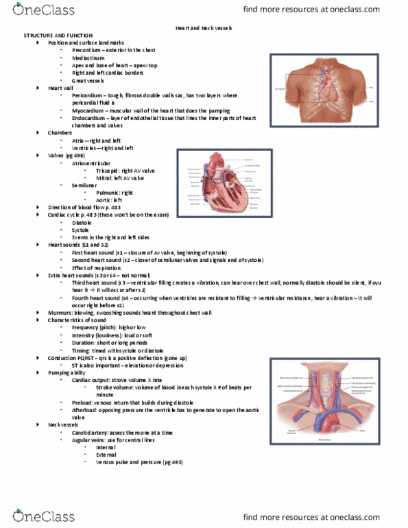
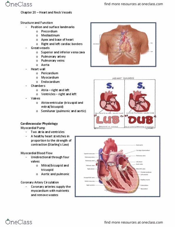
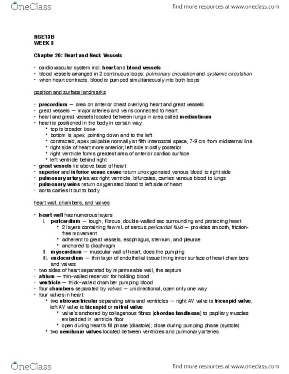
Into the right side of the heart: afterload: pressure generated in the ventricle to open the aortic valve (resistance). Position and surface landmarks: precordium external chest, anterior portion, mediastinum, apex and base of the heart apex is at the 5th intercostal space at left midclavicular line. Heart wall: pericardium - tough, fibrous sac, myocardium muscle wall of the heart, thick, endocardium thin layer that lines inner surface of the heart, serous, moist. Chambers: atria right and left, ventricles right and left. Valves: tricuspid (av valve)- right atrium and ventricle, mitral (av valve) left atria and ventricle, pulmonic (semilunar valve) separates right ventricle with pulmonary artery, aortic (semilunar valve) Vena cavae right atrium - (tricuspid valve)- right ventricle - pulmonary artery (pulmonic valve)- lungs pulmonary vein left atrium - (mitral valve) - left ventricle . Pulmonary artery is the only artery with venous blood.