NURS 301 Lecture 5: Online Lecture - Week 5 (Brain Anatomy)


Brain and Cranial Nerves
We are born with disproportionate sized brain
Baby head is 50% of weight in baby body
○
Adult is 2-3% of weight in adult body
○
Vessels and plates are more fragile
Less myelination as a baby
§
○
Baby brain more susceptible to trauma and damage
because of soft spots
○
-
Adult Brain
Average weight is 3 lbs
○
Receives 20% of blood vessels
Going through major vessels (carotid arteries)
§
○
The way we define death has to do with the state of our
brain
Death = lack of all brain activity
§
○
-
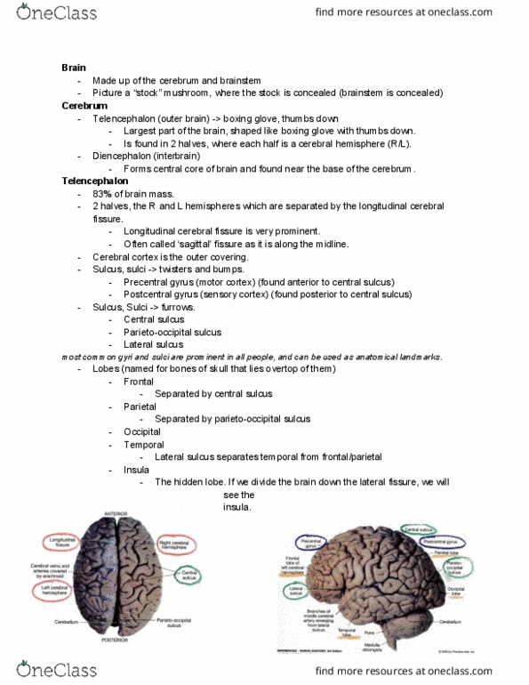
Surface features (MAJOR LANDMARKS)
Cerebrum
Cerebral hemispheres
§
Longitudinal fissure
§
Not a smooth surface
Has bounds and deep valleys
□
Gyri (Gyrus) = mounds
□
Sulci (Sulcus) = deep depressions
□
§
Gyri and sulci increase surface area
§
○
Inferior is CEREBELLUM
○
Brainstem = primitive brain
○
-
Cutting in the brain
Gray and white matter
Gray is OUTSIDE
Cell bodies, dendrites
□
Synapsing is occurring
□
Nuclei = clusters of cell bodies
□
§
White is INSIDE
Myelinated axons running through the brain
□
Tracts
□
§
○
-
Spinal Cord
Gray inside
○
White outside
○
-
Protection of Brain
Cranium
Bony
○
-
Cranial meninges
Layer of protection
○
Dura mater - tough, outer layer
Splits into two layers
§
Forming vessels called sinuses
Dural sinuses
□
§
Longitudinal fissure
Separates division between two halves
□
§
○
Arachnoid mater - transparent, middle layer
○
Pia mater - delicate, inner layer
○
-
Cerebral spinal fluid
Bathes the brain and found in spaces within the brain
○
Ventricles: chambers filled with CSF
○
Lateral look like horns
Connects to third ventricle on brain stem
§
○
Diamond shape across from cerebellum
○
Similar to the fluid portion of blood (plasma)
Salty solution
§
Greater H+
§
Less K+
§
○
No blood cells or large proteins
○
Exchange of solutes between CNS interstitial fluid and
CSF
○
Spinal Tap?
Lumbar puncture
§
Done to determine if there's an infection
§
○
-
Blood-brain barrier
-
How is CSF produced?
Produced in Choroid Plexuses of third ventricle
Finger like projections coming down from roof of
ventricles
○
-
Specially designed capillaries
Pia mater around it
○
Outside is ependymal cells
○
-
Capillaries are leaky
Ependymal cells are controlling what is going through and
filter
○
Very carefully monitored
○
Only certain material can pass through
○
-
Cerebral Spinal Fluid Circulation and Absorption into the
Blood via the Arachnoid Granulations
CSF produced in spaces
-
Transported around the brain and in spinal cord
-
And in central canal of spinal cord
-
Specific route it flows through
-
Starting from lateral ventricles (ram horns)
Goes through foramen through third ventricles
○
Picks up more CSF
○
Passes through canal, cerebral aqueduct
○
Goes through 4th ventricle
○
Passes through lateral and medial aperture
Where it flows through subarachnoid space
§
○
Excess is delivered back into blood supply
○
-
Make about 500 mL of CSF a day
Buildup of CSF is bad
○
Always must make fresh and drain away
○
Must deliver back into blood supply
○
-
Hydrocephalus
Obstruction of CSF circulation or drainage
-
Could have tumors that block drainage
-
Damage to tissues that obscures flow of CSF
-
Narrowing of aqueduct
-
LEADS TO BUILD UP
-
Halter Valve
Drains CSF
○
-
Blood-Brain Barrier
Brains require a lot of blood, 20% of blood goes to the brain
-
Requires a lot of oxygen and glucose
-
Brain is very sensitive to chemicals or microorganisms that
could penetrate it
-
Brain does not have a strong defense system
-
Design is link with capillary
Endothelial cells connected
○
Materials must be delivered to the brain microvessel
○
-
Parkinson's Disease
Lost of neurons that produce dopamine (neurotransmitter)
○
Dopamine cannot be delivered in the blood because it does
not pass the blood brain barrier
○
L-Dopa can be delivered to the brain, but limited value for
treatment
○
-
Benadryl (sleepy anti-histamine)
Sleepy because it passes through the blood-brain barrier
○
-
Major Regions of the Brain
Overview of the brain
Primitive brain: brainstem
○
Fore brain (superior regions of brain)
Diencephalon
§
Cerebrum
§
○
-
Brainstem
Going up from the spinal cord
Medulla
Starts at foramen magnum
□
Between spinal cord and the pons
□
Cross section of Medulla
Tracts: myelinated axons that carry
messages through the central nervous
system
®
Nuclei: clusters of cell bodies of the
neurons
Control many involuntary functions
Blood pressure
}
Respiratory center
}
Cardiac center
}
◊
®
Pyramids: long bulbous structure
Areas where tracts decussate
Going from right to left or left to
right
}
◊
®
□
Cranial nerves enter the medulla, five of them
□
§
Pons
Between medulla and midbrain
□
Cross section of Pons
Pons means bridge
®
Connects cerebrum to cerebellum
Cerebellum: inferior and posterior
◊
®
□
Important relay information between cerebrum
and cerebellum
□
Coordination of breathing rate
□
Involuntary visceral and somatic control of
muscles and internal organs
□
4 pairs of cranial nerves enter Pons
□
Respiration, sleep, bladder control
□
§
Midbrain
Between Pons and Diencephalon
□
Cross Section of Midbrain
Control visual tracking, following visual
view
®
Auditory and visual reflexes
®
Important reflex centers
®
□
Substantia nigra
Appears to relay inhibitory signals to the
thalamus
®
Suppresses unwanted movements so you
don't have excess or tremor, shaking like
movements
®
Important with Parkinson's disease
®
□
Copora Quadrigemina "4 bodies:
Superior and inferior colliculi
Superior are associated with visual
reflexes
◊
Inferior associated with auditory
◊
®
□
§
○
-
Diencephalon: between the Midbrain and Cerebrum
Major portions: thalamus, hypothalamus, pineal gland
○
Beneath hypothalamus is pituitary gland (sits on
sphenoid bone)
○
Largest portion is thalamus
Principle relay stations of information to our upper
brain and cerebral cortex area
§
Synapse of information coming from spinal cord and
going to cerebral cortex
§
Integration center
Knowledge acquisition
□
Cognition
□
Recognition
□
§
○
Hypothalamus
Homeostatic centers
How much food you take in
□
Temperature
□
Cardiovascular control
□
Bladder
□
Digestive
□
§
Involuntary control
§
Influence emotional state
§
○
Pituitary Gland
Hormones from hypothalamus has influence on gland
§
○
Pineal Gland
Secretes hormone melatonin
Sleep aid
□
Induces sleep
□
Darkness hormone
□
Seasonal Depression Disorder
□
§
○
-
The Reticular Formation
Web of gray matter that runs through all levels of brainstem
-
Functions
Somatic motor control
○
Cardiovascular control
○
Pain modulation
○
Sleep and consciousness
○
Habituation
Ignoring inconsequentially stimulus
§
Ignores unimportant information
Ex: TV in next room, clock ticking
□
§
○
-
Clusters of cell bodies scattered throughout the brain stem
-
Cyclical motor functions
Rhythmic activities
Breathing in and out
§
Walking
§
Chewing
§
○
-
Linked with cardiovascular center of medulla
Deals with heartrate
○
-
Cerebellum "little brain"
Second largest part of the brain
-
Muscular balance and control
-
Receives information from somatic receptors and receptors of
equilibrium and balance in the inner ear; also motor input from
cerebrum
-
Has 2 hemispheres
Vermis connects the two halves
○
-
Arbor vitae "tree of life"
-
Principal pathways of cerebellar
As you use muscles, you are getting information of what's
happening
○
Getting sensory input from vision and ear for balance and
equilibrium, continuous adjustment of movement
○
Efficient and smooth movement from cerebrum
○
-
Involved with language and cognition
-
Emotional role, spatial perception,
-
Cerebrum
Largest portion of the brain, superior portion of the brain
-
Highest brain activity and functions
Thought
○
Judgement
○
Memory
○
Comprehension
○
-
Gray matter on the outside, makes up cerebral cortex
-
Tracts running in white matter
-
Longitudinal fissure
Divides two cerebral hemispheres
○
-
Deep in the fissure
Corpus callosum
Band of white matter
§
Contains tracts
§
Carries information between the hemispheres
§
○
-
Tracts in cerebrum cross in the corpus callosum so that you
exchange information between two hemispheres
-
Tracts connecting between the same hemispheres as well
-
Lobes of Cerebrum
Frontal
anterior
§
○
Parietal
Right and left
§
○
Occipital
posterior
§
○
Temporal
Right and left
§
Pull back the temporal lobe, there is insula
§
○
-
Cerebral Cortex Functional Areas
Sensory areas: process sensations
Primary somatic: temperature, touch, pain, itch, body
position (parietal lobe)
○
Visual: sight (occipital lob)
○
Auditory: sound (temporal)
○
Olfactory: smell (temporal and frontal)
○
Gustatory: taste (insula and parietal)
○
-
Motor areas: initiate movement
-
Association areas: integration
Adjacent to sensory areas; further analysis
○
Complex integration functions
Memory
§
Emotion
§
Reasoning
§
Will
§
Judgement
§
Personality
§
Intelligence
§
○
-
The primary somatosensory area (postcentral gyrus)
Postcentral gyrus contains primary somatosensory cortex
-
Exhibits somatotopy - a body map
Specific to particular body areas
○
-
Central sulcus
-
Precentral gyrus
Motor activity
○
-
Language is one of the hallmarks of an advanced nervous
system:
Language skills require the input of sensory info (mainly
hearing and vision), processing, and coordination of motor
output for writing and vocalization
-
In most people the integration of spoken language in the human
brain is the left hemisphere of the cerebral cortex
Two major cortex areas involves
Wernicke's area: association area; interpretation of
speech, translation of words to though
Parietal area
□
Primary auditory cortex
□
§
Broca's area: motor area; production of speech;
formation of words
Frontal area
□
Premotor area
□
§
○
Aphasia: language problem
Could be due to interpretation or production of speech
§
Ability to write
§
○
-
PET Scans of the Brain during performance of a language
task
The word car is seen in the visual cortex
Occipital lobe
-
Sensory input from out eyes
-
1.
Wernicke area conceives off the verb DRIVE to go with it2.
Broca area compiles a motor program to speak the word
DRIVE
3.
Primary motor cortex executes the program and the word
is spoken
4.
-
Lateralization of Cerebral Functions
Difference in functions for each hemisphere
-
Categorical hemisphere
-
Representational hemisphere
-
Right controls left
-
Left controls right
-
Left hemisphere
Controls language, numerical, scientific skills sign
language, reasoning
○
-
Right hemisphere
Creative side of brain, music, artistic, spatial and pattern
perception, emotional intent, content of language
○
-
Limbic System
Prominent parts include: cingulate gyrus, hippocampus,
amygdala
-
Important functions for emotion and learning
-
Contains multiple gratification and aversion centers
-
Involved in processing of emotion
Emotion is linked to learn
○
Involved with our cognition
○
-
Cognition, Memory
Cognition
Acquire and use knowledge
○
Association areas of cortex
Synapses occur in these areas
Induces a sustain change
□
As you practice something, quality improves
□
Able to remember cranial nerves
□
-
○
-
Memory
Two types: procedural and declarative
Procedural: repeated process that you do after awhile
without thinking
Riding a bike, playing an instrument, texting
□
You have memory for these, networks allow us
to do it
□
-
Declarative
Memory of a name or someone's birthday
□
-
○
Limbic areas involved
Amygdala creates emotional memories
-
Hippocampus consolidates declarative long-term
memories
-
○
-
Alzheimer's Disease Brain
Degenerative progressive disease
Gets worse with time
○
-
Characteristic memory loss
Portions of cerebral cortex loss
○
Ex: forget how to get home, forget loved ones names and
faces
○
-
Plaques and tangles
-
Diagnostic tool
Electroencephalogram (EEG)
Special electrodes on scalp on patient
Little hair as possible
□
-
Neuronal activity at surface of head
-
Read waveforms
Alpha waves: characteristic of normal resting
adult
□
Beta waves: typically accompany intense
concentration
□
Theta waves: seen in children and in frustrated
adults
□
Delta waves: occur in deep sleeve and in certain
pathological states
Alzheimer's patient
®
□
-
○
-
Cranial Nerves
12 pairs of peripheral nerves
-
Each pair
Identified by roman numeral and name
○
I enters through the forebrain
○
II through XII originate along the brain stem
○
Sensory, motor, or mixed nerves
Ex: Vagus, X, is a mixed nerve to many internal
organs, muscles, and glands
Extends from medulla, enters brain and into
jugular foramen (opening in cranium)
□
Goes down into neck and thoracic area
□
-
○
Pairs on each side
○
-
Cranial nerves have branching patterns into face
-
Online Lecture - Week 5 (Brain Anatomy)
Wednesday, April 25, 2018
1:57 PM

Brain and Cranial Nerves
We are born with disproportionate sized brain
Baby head is 50% of weight in baby body
○
Adult is 2-3% of weight in adult body
○
Vessels and plates are more fragile
Less myelination as a baby
§
○
Baby brain more susceptible to trauma and damage
because of soft spots
○
-
Adult Brain
Average weight is 3 lbs
○
Receives 20% of blood vessels
Going through major vessels (carotid arteries)
§
○
The way we define death has to do with the state of our
brain
Death = lack of all brain activity
§
○
-
Surface features (MAJOR LANDMARKS)
Cerebrum
Cerebral hemispheres
§
Longitudinal fissure
§
Not a smooth surface
Has bounds and deep valleys
□
Gyri (Gyrus) = mounds
□
Sulci (Sulcus) = deep depressions
□
§
Gyri and sulci increase surface area
§
○
Inferior is CEREBELLUM
○
Brainstem = primitive brain
○
-
Cutting in the brain
Gray and white matter
Gray is OUTSIDE
Cell bodies, dendrites
□
Synapsing is occurring
□
Nuclei = clusters of cell bodies
□
§
White is INSIDE
Myelinated axons running through the brain
□
Tracts
□
§
○
-
Spinal Cord
Gray inside
○
White outside
○
-
Protection of Brain
Cranium
Bony
○
-
Cranial meninges
Layer of protection
○
Dura mater - tough, outer layer
Splits into two layers
§
Forming vessels called sinuses
Dural sinuses
□
§
Longitudinal fissure
Separates division between two halves
□
§
○
Arachnoid mater - transparent, middle layer
○
Pia mater - delicate, inner layer
○
-
Cerebral spinal fluid
Bathes the brain and found in spaces within the brain
○
Ventricles: chambers filled with CSF
○
Lateral look like horns
Connects to third ventricle on brain stem
§
○
Diamond shape across from cerebellum
○
Similar to the fluid portion of blood (plasma)
Salty solution
§
Greater H+
§
Less K+
§
○
No blood cells or large proteins
○
Exchange of solutes between CNS interstitial fluid and
CSF
○
Spinal Tap?
Lumbar puncture
§
Done to determine if there's an infection
§
○
-
Blood-brain barrier
-
How is CSF produced?
Produced in Choroid Plexuses of third ventricle
Finger like projections coming down from roof of
ventricles
○
-
Specially designed capillaries
Pia mater around it
○
Outside is ependymal cells
○
-
Capillaries are leaky
Ependymal cells are controlling what is going through and
filter
○
Very carefully monitored
○
Only certain material can pass through
○
-
Cerebral Spinal Fluid Circulation and Absorption into the
Blood via the Arachnoid Granulations
CSF produced in spaces
-
Transported around the brain and in spinal cord
-
And in central canal of spinal cord
-
Specific route it flows through
-
Starting from lateral ventricles (ram horns)
Goes through foramen through third ventricles
○
Picks up more CSF
○
Passes through canal, cerebral aqueduct
○
Goes through 4th ventricle
○
Passes through lateral and medial aperture
Where it flows through subarachnoid space
§
○
Excess is delivered back into blood supply
○
-
Make about 500 mL of CSF a day
Buildup of CSF is bad
○
Always must make fresh and drain away
○
Must deliver back into blood supply
○
-
Hydrocephalus
Obstruction of CSF circulation or drainage
-
Could have tumors that block drainage
-
Damage to tissues that obscures flow of CSF
-
Narrowing of aqueduct
-
LEADS TO BUILD UP
-
Halter Valve
Drains CSF
○
-
Blood-Brain Barrier
Brains require a lot of blood, 20% of blood goes to the brain
-
Requires a lot of oxygen and glucose
-
Brain is very sensitive to chemicals or microorganisms that
could penetrate it
-
Brain does not have a strong defense system
-
Design is link with capillary
Endothelial cells connected
○
Materials must be delivered to the brain microvessel
○
-
Parkinson's Disease
Lost of neurons that produce dopamine (neurotransmitter)
○
Dopamine cannot be delivered in the blood because it does
not pass the blood brain barrier
○
L-Dopa can be delivered to the brain, but limited value for
treatment
○
-
Benadryl (sleepy anti-histamine)
Sleepy because it passes through the blood-brain barrier
○
-
Major Regions of the Brain
Overview of the brain
Primitive brain: brainstem
○
Fore brain (superior regions of brain)
Diencephalon
§
Cerebrum
§
○
-
Brainstem
Going up from the spinal cord
Medulla
Starts at foramen magnum
□
Between spinal cord and the pons
□
Cross section of Medulla
Tracts: myelinated axons that carry
messages through the central nervous
system
®
Nuclei: clusters of cell bodies of the
neurons
Control many involuntary functions
Blood pressure
}
Respiratory center
}
Cardiac center
}
◊
®
Pyramids: long bulbous structure
Areas where tracts decussate
Going from right to left or left to
right
}
◊
®
□
Cranial nerves enter the medulla, five of them
□
§
Pons
Between medulla and midbrain
□
Cross section of Pons
Pons means bridge
®
Connects cerebrum to cerebellum
Cerebellum: inferior and posterior
◊
®
□
Important relay information between cerebrum
and cerebellum
□
Coordination of breathing rate
□
Involuntary visceral and somatic control of
muscles and internal organs
□
4 pairs of cranial nerves enter Pons
□
Respiration, sleep, bladder control
□
§
Midbrain
Between Pons and Diencephalon
□
Cross Section of Midbrain
Control visual tracking, following visual
view
®
Auditory and visual reflexes
®
Important reflex centers
®
□
Substantia nigra
Appears to relay inhibitory signals to the
thalamus
®
Suppresses unwanted movements so you
don't have excess or tremor, shaking like
movements
®
Important with Parkinson's disease
®
□
Copora Quadrigemina "4 bodies:
Superior and inferior colliculi
Superior are associated with visual
reflexes
◊
Inferior associated with auditory
◊
®
□
§
○
-
Diencephalon: between the Midbrain and Cerebrum
Major portions: thalamus, hypothalamus, pineal gland
○
Beneath hypothalamus is pituitary gland (sits on
sphenoid bone)
○
Largest portion is thalamus
Principle relay stations of information to our upper
brain and cerebral cortex area
§
Synapse of information coming from spinal cord and
going to cerebral cortex
§
Integration center
Knowledge acquisition
□
Cognition
□
Recognition
□
§
○
Hypothalamus
Homeostatic centers
How much food you take in
□
Temperature
□
Cardiovascular control
□
Bladder
□
Digestive
□
§
Involuntary control
§
Influence emotional state
§
○
Pituitary Gland
Hormones from hypothalamus has influence on gland
§
○
Pineal Gland
Secretes hormone melatonin
Sleep aid
□
Induces sleep
□
Darkness hormone
□
Seasonal Depression Disorder
□
§
○
-
The Reticular Formation
Web of gray matter that runs through all levels of brainstem
-
Functions
Somatic motor control
○
Cardiovascular control
○
Pain modulation
○
Sleep and consciousness
○
Habituation
Ignoring inconsequentially stimulus
§
Ignores unimportant information
Ex: TV in next room, clock ticking
□
§
○
-
Clusters of cell bodies scattered throughout the brain stem
-
Cyclical motor functions
Rhythmic activities
Breathing in and out
§
Walking
§
Chewing
§
○
-
Linked with cardiovascular center of medulla
Deals with heartrate
○
-
Cerebellum "little brain"
Second largest part of the brain
-
Muscular balance and control
-
Receives information from somatic receptors and receptors of
equilibrium and balance in the inner ear; also motor input from
cerebrum
-
Has 2 hemispheres
Vermis connects the two halves
○
-
Arbor vitae "tree of life"
-
Principal pathways of cerebellar
As you use muscles, you are getting information of what's
happening
○
Getting sensory input from vision and ear for balance and
equilibrium, continuous adjustment of movement
○
Efficient and smooth movement from cerebrum
○
-
Involved with language and cognition
-
Emotional role, spatial perception,
-
Cerebrum
Largest portion of the brain, superior portion of the brain
-
Highest brain activity and functions
Thought
○
Judgement
○
Memory
○
Comprehension
○
-
Gray matter on the outside, makes up cerebral cortex
-
Tracts running in white matter
-
Longitudinal fissure
Divides two cerebral hemispheres
○
-
Deep in the fissure
Corpus callosum
Band of white matter
§
Contains tracts
§
Carries information between the hemispheres
§
○
-
Tracts in cerebrum cross in the corpus callosum so that you
exchange information between two hemispheres
-
Tracts connecting between the same hemispheres as well
-
Lobes of Cerebrum
Frontal
anterior
§
○
Parietal
Right and left
§
○
Occipital
posterior
§
○
Temporal
Right and left
§
Pull back the temporal lobe, there is insula
§
○
-
Cerebral Cortex Functional Areas
Sensory areas: process sensations
Primary somatic: temperature, touch, pain, itch, body
position (parietal lobe)
○
Visual: sight (occipital lob)
○
Auditory: sound (temporal)
○
Olfactory: smell (temporal and frontal)
○
Gustatory: taste (insula and parietal)
○
-
Motor areas: initiate movement
-
Association areas: integration
Adjacent to sensory areas; further analysis
○
Complex integration functions
Memory
§
Emotion
§
Reasoning
§
Will
§
Judgement
§
Personality
§
Intelligence
§
○
-
The primary somatosensory area (postcentral gyrus)
Postcentral gyrus contains primary somatosensory cortex
-
Exhibits somatotopy - a body map
Specific to particular body areas
○
-
Central sulcus
-
Precentral gyrus
Motor activity
○
-
Language is one of the hallmarks of an advanced nervous
system:
Language skills require the input of sensory info (mainly
hearing and vision), processing, and coordination of motor
output for writing and vocalization
-
In most people the integration of spoken language in the human
brain is the left hemisphere of the cerebral cortex
Two major cortex areas involves
Wernicke's area: association area; interpretation of
speech, translation of words to though
Parietal area
□
Primary auditory cortex
□
§
Broca's area: motor area; production of speech;
formation of words
Frontal area
□
Premotor area
□
§
○
Aphasia: language problem
Could be due to interpretation or production of speech
§
Ability to write
§
○
-
PET Scans of the Brain during performance of a language
task
The word car is seen in the visual cortex
Occipital lobe
-
Sensory input from out eyes
-
1.
Wernicke area conceives off the verb DRIVE to go with it2.
Broca area compiles a motor program to speak the word
DRIVE
3.
Primary motor cortex executes the program and the word
is spoken
4.
-
Lateralization of Cerebral Functions
Difference in functions for each hemisphere
-
Categorical hemisphere
-
Representational hemisphere
-
Right controls left
-
Left controls right
-
Left hemisphere
Controls language, numerical, scientific skills sign
language, reasoning
○
-
Right hemisphere
Creative side of brain, music, artistic, spatial and pattern
perception, emotional intent, content of language
○
-
Limbic System
Prominent parts include: cingulate gyrus, hippocampus,
amygdala
-
Important functions for emotion and learning
-
Contains multiple gratification and aversion centers
-
Involved in processing of emotion
Emotion is linked to learn
○
Involved with our cognition
○
-
Cognition, Memory
Cognition
Acquire and use knowledge
○
Association areas of cortex
Synapses occur in these areas
Induces a sustain change
□
As you practice something, quality improves
□
Able to remember cranial nerves
□
-
○
-
Memory
Two types: procedural and declarative
Procedural: repeated process that you do after awhile
without thinking
Riding a bike, playing an instrument, texting
□
You have memory for these, networks allow us
to do it
□
-
Declarative
Memory of a name or someone's birthday
□
-
○
Limbic areas involved
Amygdala creates emotional memories
-
Hippocampus consolidates declarative long-term
memories
-
○
-
Alzheimer's Disease Brain
Degenerative progressive disease
Gets worse with time
○
-
Characteristic memory loss
Portions of cerebral cortex loss
○
Ex: forget how to get home, forget loved ones names and
faces
○
-
Plaques and tangles
-
Diagnostic tool
Electroencephalogram (EEG)
Special electrodes on scalp on patient
Little hair as possible
□
-
Neuronal activity at surface of head
-
Read waveforms
Alpha waves: characteristic of normal resting
adult
□
Beta waves: typically accompany intense
concentration
□
Theta waves: seen in children and in frustrated
adults
□
Delta waves: occur in deep sleeve and in certain
pathological states
Alzheimer's patient
®
□
-
○
-
Cranial Nerves
12 pairs of peripheral nerves
-
Each pair
Identified by roman numeral and name
○
I enters through the forebrain
○
II through XII originate along the brain stem
○
Sensory, motor, or mixed nerves
Ex: Vagus, X, is a mixed nerve to many internal
organs, muscles, and glands
Extends from medulla, enters brain and into
jugular foramen (opening in cranium)
□
Goes down into neck and thoracic area
□
-
○
Pairs on each side
○
-
Cranial nerves have branching patterns into face
-
Online Lecture - Week 5 (Brain Anatomy)
Wednesday, April 25, 2018 1:57 PM
Document Summary
Baby head is 50% of weight in baby body. Adult is 2-3% of weight in adult body. Baby brain more susceptible to trauma and damage because of soft spots. The way we define death has to do with the state of our brain. Bathes the brain and found in spaces within the brain. Similar to the fluid portion of blood (plasma) Exchange of solutes between cns interstitial fluid and. Finger like projections coming down from roof of ventricles. Ependymal cells are controlling what is going through and filter. Cerebral spinal fluid circulation and absorption into the. Transported around the brain and in spinal cord. Make about 500 ml of csf a day. Damage to tissues that obscures flow of csf. Brains require a lot of blood, 20% of blood goes to the brain. Brain is very sensitive to chemicals or microorganisms that could penetrate it. Brain does not have a strong defense system.