BCS 151 Lecture Notes - Lecture 12: Biological Motion, Optical Flow, Akinetopsia

20 Oct 2016
School
Department
Course
Professor
Document Summary
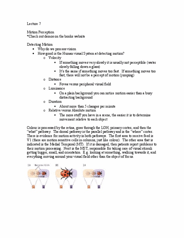
Motion detection takes place in the retina, lgn and v1. Interpretation of motion takes place in the mt and mst regions. In v1, motion is detected by directional selective complex cells (~15%) Apparent motion if you flash something back and forth, even there is no continuous motion, you still detect the object as moving. Fast motion the m cells on the above diagram as much further apart: more of a delay is necessary before the signals align and produce a coincidence. 1 & 2/3&4 the image goes forward. 2 & 3/1&4 the image goes backwards. Area of the m pathway critical for normal motion perception. Mt has many interconnections with other regions of the cortex. Located in the back of the brain (occipital cortex) The receptive field is 10x greater than what is in the v1: many to one convergence from the neurons in the v1 cortex, gives the overall object, instead of its parts, where integration occurs!