ANA 407 Lecture Notes - Lecture 14: Esophageal Hiatus, Azygos Vein, Thoracic Cavity

8 Jun 2016
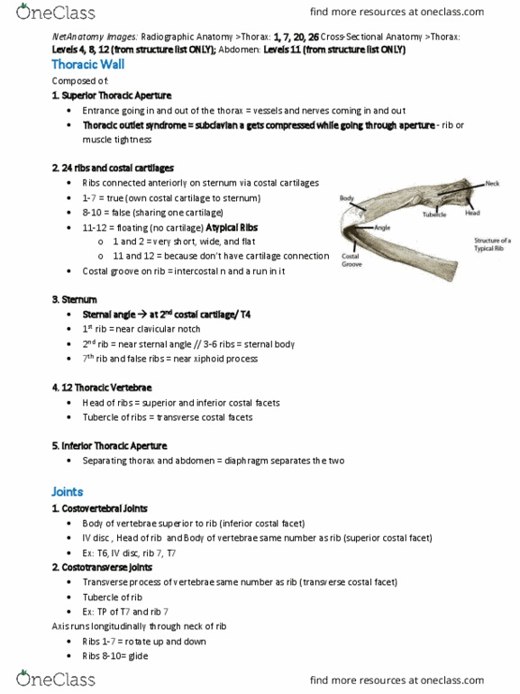
School
Department
Course
Professor
Document Summary
Describe the diaphragm and its anatomical relationships. a. thoracic. The diaphragm is a musculotendinous sheet that separates the and abdominal cavities. The muscle of the diaphragm is skeletal muscle, and is under voluntary control, but it is also driven automatically by respiratory reflexes. It is divided into three parts that insert onto the central tendon: 2) sternal part - arises from the xiphoid process costal part - from the internal surfaces of the lower. 6 ribs, at the costal margin lumbar part - from lumbar vertebrae as low as l3 on the right, and l2 on the left. Note that fibers of the right crus encircle the esophageal hiatus. The major structures that pass through or behind the diaphragm include the: 1) descending aorta - passes behind the diaphragm at the junction of the left and right crura. The aortic hiatus is the gap through which the aorta passes, usually in front of the t12 vertebral body.