BSC 310 Lecture Notes - Lecture 5: Malachite Green, Gram Staining, Methylene Blue


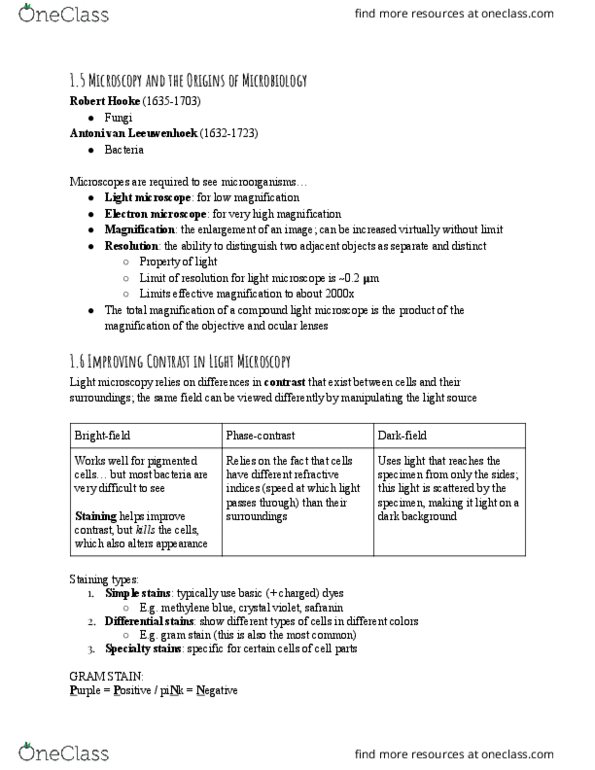
Staining Techniques
Because microbial cytoplasm is usually transparent, it is necessary to stain
microorganisms before they can be viewed with the light microscope. In some cases,
staining is unnecessary, for example when microorganisms are very large or when
motility is to be studied, and a drop of the microorganisms can be placed directly on the
slide and observed. A preparation such as this is called a wet mount. A wet mount can
also be prepared by placing a drop of culture on a cover slip (a glass cover for a slide) ‐
and then inverting it over a hollowed out slide. This procedure is called the‐ hanging
drop.
In preparation for staining, a small sample of microorganisms is placed on a slide and
permitted to air dry. The smear is heat fixed by quickly passing it over a flame. Heat
fixing kills the organisms, makes them adhere to the slide, and permits them to accept
the stain.
Simple stain techniques. Staining can be performed with basic dyes such as crystal
violet or methylene blue, positively charged dyes that are attracted to the negatively
charged materials of the microbial cytoplasm. Such a procedure is the simple stain
procedure. An alternative is to use a dye such as nigrosin or Congo red, acidic,
negatively charged dyes. They are repelled by the negatively charged cytoplasm and
gather around the cells, leaving the cells clear and unstained. This technique is called
the negative stain technique.
Differential stain techniques. The differential stain technique distinguishes two
kinds of organisms. An example is the Gram stain technique. This differential
technique separates bacteria into two groups, Gram positive bacteria and Gram‐ ‐
negative bacteria. Crystal violet is first applied, followed by the mordant iodine, which
fixes the stain (Figure ). Then the slide is washed with alcohol, and the Gram positive ‐
bacteria retain the crystal violet iodine stain; however, the Gram negative bacteria lose ‐ ‐
the stain. The Gram negative bacteria subsequently stain with the safranin dye, the ‐
counterstain, used next. These bacteria appear red under the oil immersion lens, while ‐
Gram positive bacteria appear blue or purple, reflecting the crystal violet retained during‐
the washing step.
Another differential stain technique is the acid fast technique.‐ This technique
differentiates species of Mycobacterium from other bacteria. Heat or a lipid solvent is
used to carry the first stain, carbolfuchsin, into the cells. Then the cells are washed with
a dilute acid alcohol solution.‐ Mycobacterium species resist the effect of the acid‐
alcohol and retain the carbolfuchsin stain (bright red). Other bacteria lose the stain and
take on the subsequent methylene blue stain (blue). Thus, the acid fast bacteria appear ‐
bright red, while the nonacid fast bacteria appear blue when observed under oil‐ ‐
immersion microscopy.
Other stain techniques seek to identify various bacterial structures of importance. For
instance, a special stain technique highlights the flagella of bacteria by coating the
flagella with dyes or metals to increase their width. Flagella so stained can then be
observed.
find more resources at oneclass.com
find more resources at oneclass.com
Document Summary
Because microbial cytoplasm is usually transparent, it is necessary to stain microorganisms before they can be viewed with the light microscope. In some cases, staining is unnecessary, for example when microorganisms are very large or when motility is to be studied, and a drop of the microorganisms can be placed directly on the slide and observed. A preparation such as this is called a wet mount. A wet mount can also be prepared by placing a drop of culture on a cover slip (a glass cover for a slide) and then inverting it over a hollowed out slide. In preparation for staining, a small sample of microorganisms is placed on a slide and permitted to air dry. The smear is heat fixed by quickly passing it over a flame. Heat fixing kills the organisms, makes them adhere to the slide, and permits them to accept the stain.